CPS, Postural Reactions, Paw Replacement...Oh My!

CPS, Postural Reactions, Paw Replacement...Oh My!

Performing CPs, or the paw replacement test, is so important for lesion localization, or to even identify if a patient has a neurologic problem. But it is difficult! This test causes residents to fret and frown because subtle differences in how you hold a patient, the flooring, and even environmental stimuli can affect how they place their limb. If you practice…it gets easier!

How do you identify increased intracranial pressures following head trauma?

How do you identify increased intracranial pressures following head trauma?

Are you ready to evaluate your patient for increased inttracranial pressure? Read on to see what simple techniques you can use to find this deadly result of traumatic brain injury. Spoiler alert - You need a stethoscope, a blood pressure measurement and a mini-neuro exam!

Caudal Occipital Malformation Syndrome in dogs

Caudal Occipital Malformation Syndrome in dogs

What is Caudal Occipital Malformation Syndrome? This malformation affects the caudal aspect of the skull which results in over crowding of the intracranial structures. If you think overcrowding in airplanes is tough, try over crowding your skull! The result…a multitude of clinical signs. Read on to see how this disease presents and is managed in dogs.

Do prazosin or diazepam help with urinary function following thoracolumbar spinal injury?

Does prazosin or diazepam work for urinary incontinence secondary to a thoracolumbar myelopathy?

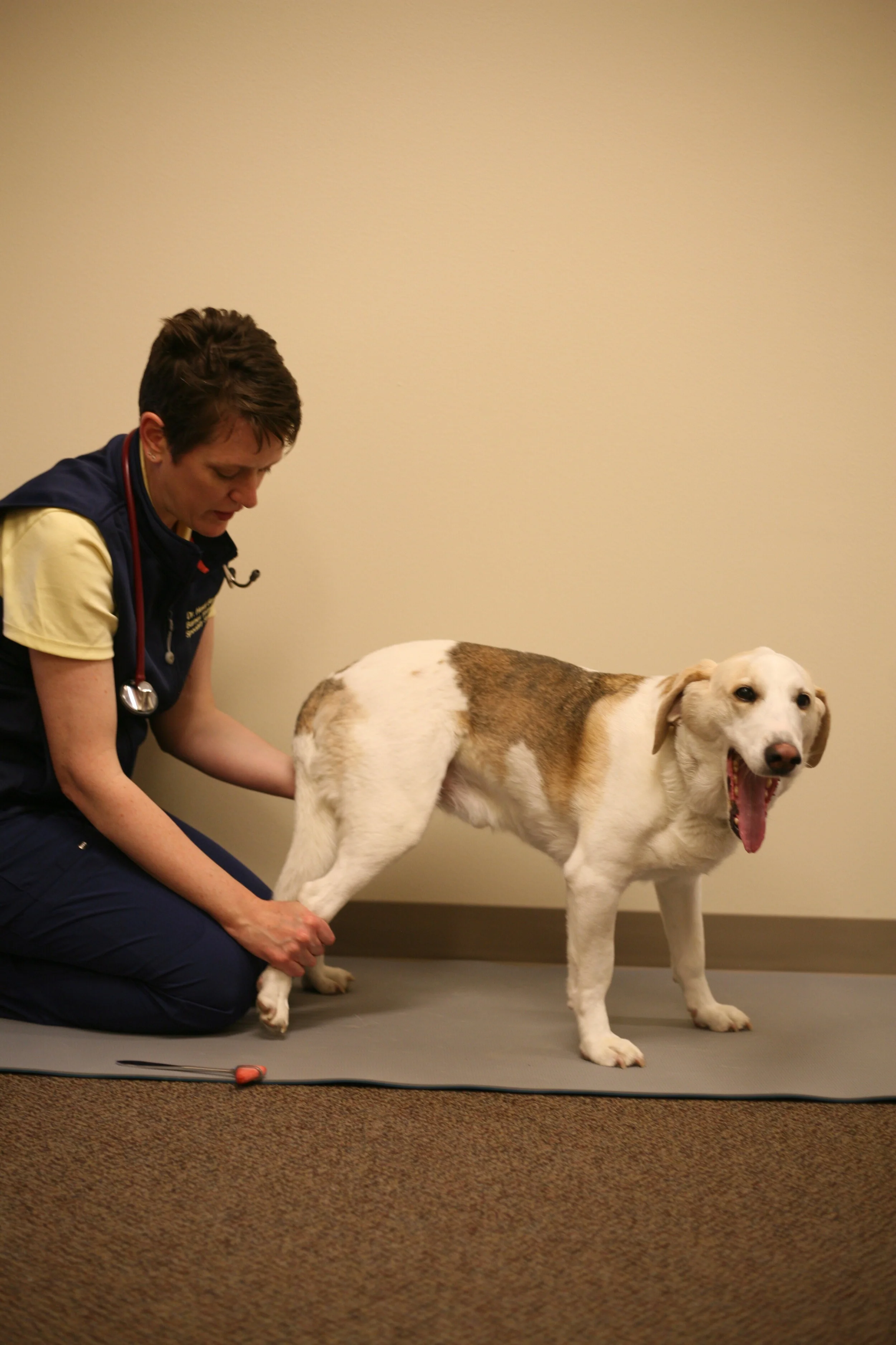
Let's back it up a moment... animals with thoracolumbar injury often have some degree of urinary incontinence if the injury is severe enough to result in a non-ambulatory status.

Here is what I expect:
No motor = no urine function.
Weak motor = +/- urine function.
Good motor = normal urine function. 

What is the neuroanatomy here?


I'm glad you asked! (If you didn't you've probably stopped reading now.) Urination is managed through a coordinated effort between the sympathetic (hypogastric nerve), parasympathetic (pelvic nerve) and somatic (pudendal nerve) function. See details below.

  1. Sympathetic innervation:

    1. Purpose: management of the storage phase of urination and pain receptors for distention in the bladder.

    2. Nerve involved: Hypogastric nerve

    3. Cell body location: L2-5 in the cat, L1-4 in the dog

    4. Neurotransmitters utilized: Beta receptors in the urine bladder and alpha receptors in the urethra are stimulated with norepinephrine to maintain a relaxed bladder muscle and tonic internal sphincter of the urethra.

** Prazosin works to block sympathetic function, thus decreasing STORAGE phase, increasing express-ability. 

  1. Parasympathetic innervation:

    1. Purpose: management of the voiding phase of urination and stretch receptors in the detrusor for distension.

    2. Nerve involved: Pelvic nerve

    3. Cell body location: S1-S3

    4. Neurotransmitters utilized: Acetylcholine is released at the nerve terminals in the bladder muscle (detrusor). There is questionable innervation of the internal sphincter with parasympathetic fibers. The most compelling research suggests that rhythmic contraction of the detrusor muscle during voiding results in stretching and opening of the urethral smooth muscle rather than direct innervation.  

  2. Somatic Innervation:

    1. Purpose: Maintenance of storage phase through contraction of the external urinary sphincter and some stretch receptors of the urethra.

    2. Nerve involved: pudendal

    3. Cell body location: S1-S3

    4. Neurotransmitters: acetylcholine

** Diazepam works here to block the external sphincter contraction.

If an animal sustains injury cranial to the pudendal/pelvic nerves (S1) they will have an "upper motor neuron" bladder. In this case, they cannot voluntarily evacuate urine. And this is when we try drugs to help these poor dogs (and owners that are trying to express them!). 

A recent article by an author with a very similar name to mine* recently published their answer to the question at the top of this mailer.


Bottom line, there was no statistical difference between the control (no drug) and drug group in terms of length of hospitalization or degree of urinary incontinence at discharge (p=0.14).


Shoot. Don't toss those drugs down the drain, however. Despite statistics, we may have some clinical improvement in individuals so it could be worth trying on a short term (7 days or less) basis. However, if it's not helping, don't continue just because it should be helping! 
 
Incidentally, they found that with each 1 kg increase in body weight the chances of incontinence increased by 14%. Yet another reason to encourage healthy-weight dachshunds!


* Barnes KH, Aulkah KS, Liu C: Retrospective evaluation of prazosin and diazepam after thoracolumbar hemilaminectomy in dogs; Vet J:253(November),2019

TidBit Tuesday 12.17.19: Degenerative Myelopathy

What is Degenerative Myelopathy?

Much more than a descriptive term, Degenerative Myelopathy (DM), is an actual disease unto itself with links to human Amyotrophic Lateral Sclerosis (ALS). 
Presenting clinical signs: pelvic limb weakness, ataxia, and paw replacement deficits 
Onset: Slow, progressive
Pain: Non-painful
Typical signalment: German Shepherd, Corgi, Boxer, Rhodesian Ridgeback most common, but other breeds possible. Typically middle aged (7+ years old) without sex predilection

Lesion localization: T3-L3 myelopathy initially, but degeneration will progress caudally causing loss of pelvic limb reflexes (in order...what is the order?) and cranially resulting in an ascending cutaneous trunci reflex and eventually loss of thoracic limb reflexes with paresis. If left to progress long enough brainstem involvement can occur. 

Diagnostic testing: 1) rule out all other causes of a T3-L3 myelopathy. These breeds are prone to IVDH type II and neoplasia both of which are slowly progressive variably painful causes of myelopathy (spinal cord disease). 2) A DNA test can be performed at several locations.I have used the Orthopedic foundation (https://www.ofa.org/diseases/dna-tested-diseases/dm) or UC Davis (https://www.vgl.ucdavis.edu/services/dog/DegenerativeMyelopathy.php) for testing. 

Interpretation of Results:
1) Not at risk: this means the dog does not have the SOD1 mutation. This mutation has been associated with the development of DM
2) Carrier: One copy of the SOD1 mutation was identified. There is a low chance of development of DM in this individual but a 50% chance of passing along one copy of the mutation should breeding occur.
3) At risk: Two copies of the SOD1 mutation have been found. This individual is at risk of developing DM. 

**Remember that a small subset of dogs can develop DM without the SOD1 mutation so necropsy is still the gold standard for a definitive diagnosis. 
 

How do I use the DNA test? 

This was a point of contention with my residents because I do not believe this test should be used in exclusion of MRI or CSF tap. I use the DM test to determine if a patient is at risk but I STILL recommend ruling out IVDH and neoplasia before I conclude that a patient actually has clinical signs attributed to DM.
 

The DNA test tells us if the dog carries one or two copies of a mutation that may or may not result in neurologic disease at some time in the dogs life. It does not tell us if the dog's current clinical signs are related to DM. 


Case in Point: An 8 year old MC Boxer presents with a 2 month history of slowly progressive difficulty walking in the pelvic limbs. On examination you localize the problem to the T3-L3 segment without evidence of spinal pain. Your differentials include: DM, neoplasia, meningomyelitis and IVDH. You do the DNA test and the dog is "at risk". What do you do? 

Option 1: Treat the dog for DM including exercise restrictions, physical rehabilitation and planning for supportive measures. No true treatment for the disease exists.

Option 2: You send them for MRI and CSF tap and a extramedullary spinal cord compression is found at T12-T13 consistent with a disc rupture. What do you do? Should the owner do surgery or not? 

In my opinion, the owner is only able to make the best decision for their pet when provided ALL of the available information. This includes doing a DNA test AND doing the MRI/CSF tap. After putting all of these pieces together we can choose a best course of action.

If we do the MRI/CSF tap first and no disease is identified do we need to do the DNA test (if not planning on breeding)? 

If the DNA test shows the patient is not at risk, does it change the fact that signs are consistent with DM and the dog has no evidence of compression or inflammatory disease? We would still treat for presumptive DM so how does the DNA test change this?

Food for thought during this season of consumption!