"Back Case" Coming In
Is anyone else exhausted after surviving May?? We haven't practiced lesion localization for a while so I thought this might be a nice easy topic for the first week of June.
History:
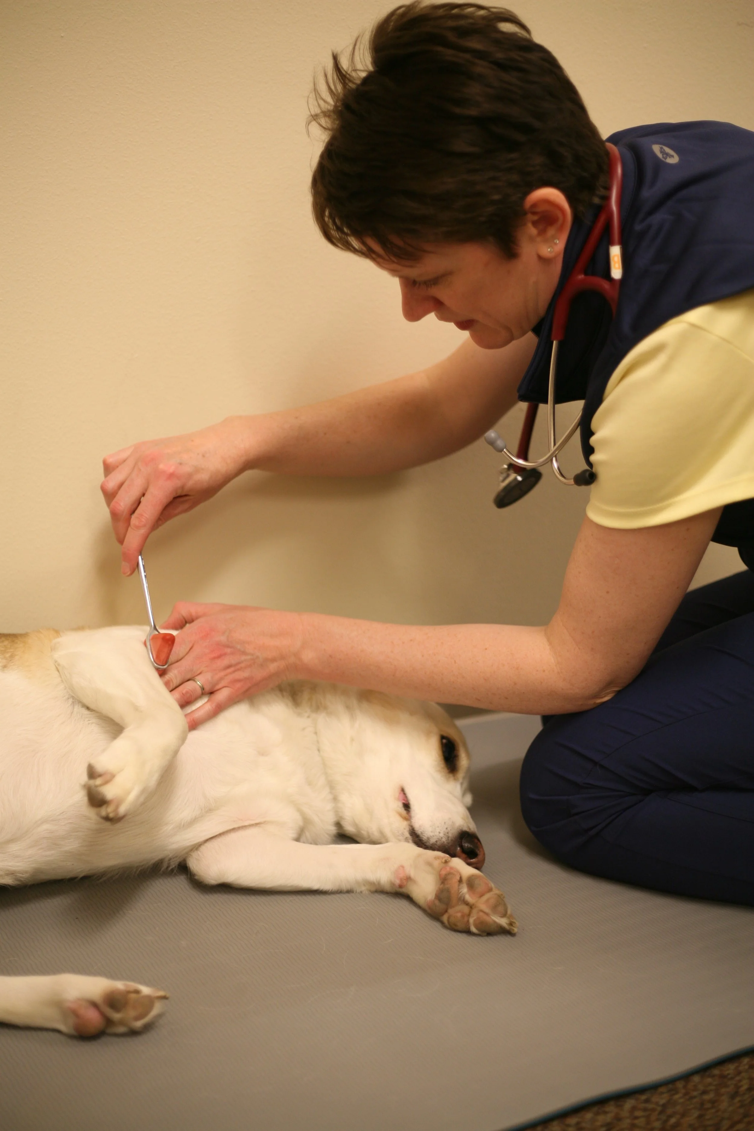
Dora is a 4 year old FS Beagle-X. She is presenting with a 7 day history of difficulty walking in the pelvic limbs with swaying, falling, and occasional vocalization in suspected pain. No prior medical history and normal physical examination.
Neurologic examination:
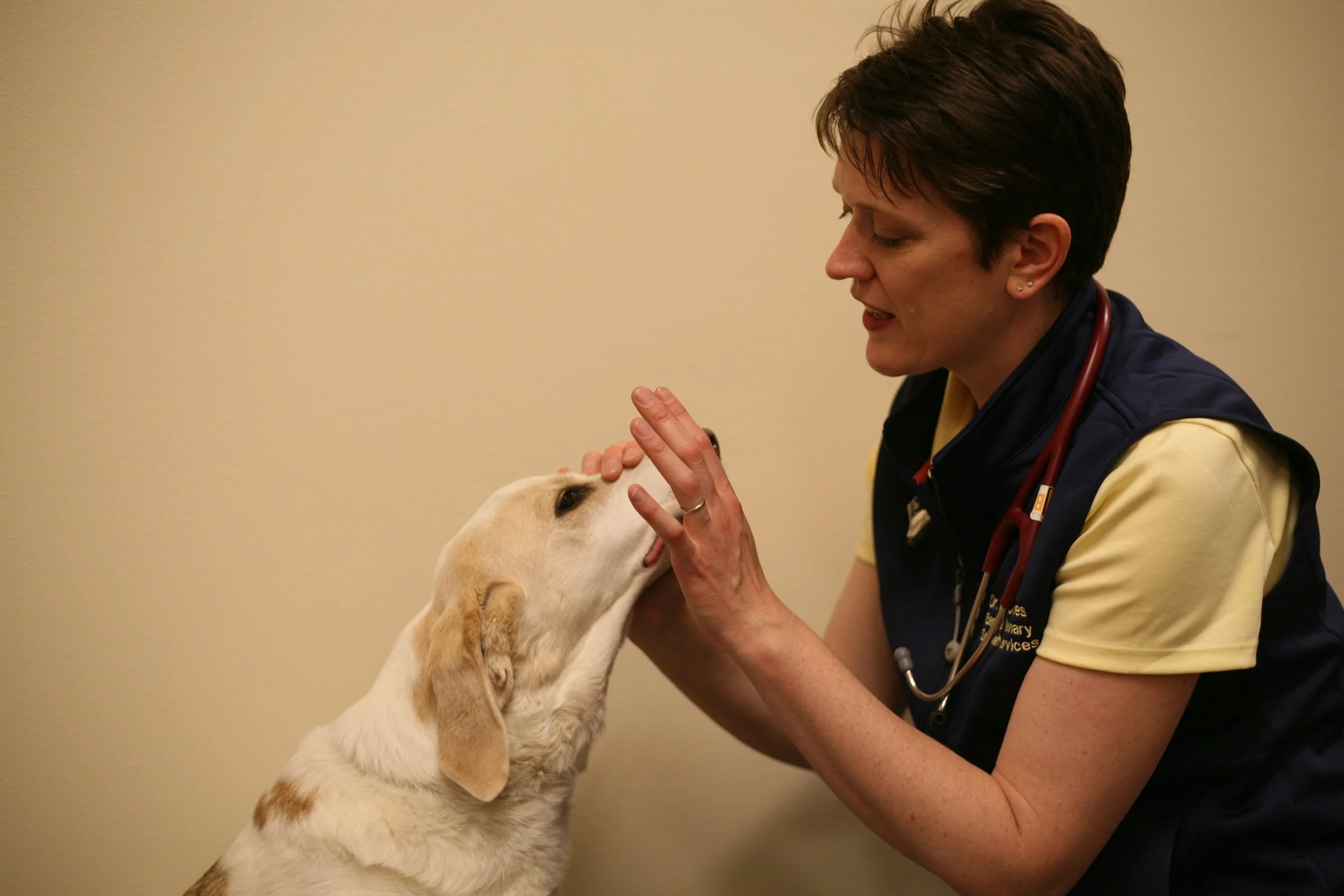
Mentation: BAR, anxious
Cranial nerves: normal
Gait: Ambulatory, paraparesis with moderate proprioceptive ataxia in pelvic limbs only.
Reflexes: normal withdrawal in all four limbs, normal patellar reflexes bilaterally and normal anal reflex. We didn't test the remainder of the limb reflexes due to her spinal pain and anxiety in lateral. The cutaneous trunci reflex stops at L2 bilaterally.
Postural reactions: Absent paw replacement testing in both pelvic limbs, normal in both thoracic limbs. Normal hopping in both thoracic limbs, absent hopping in both pelvic limbs.
Palpation: Spinal pain at TL junction, the remainder was non-painful. Normal cervical ROM and tail ROM.
The first questions we ask ourselves is "is this dog neurologically normal or abnormal?"
The answer, of course, is abnormal, so let's break it down.
Start with what you know:
This dog has normal mentation, no cranial nerve deficits and no history of behavior changes or seizures so I think we can safely assume the lesion is NOT intracranial. This leaves spinal cord, peripheral nerve, neuromuscular junction, or muscle to choose from. Here are some key points:
1. Peripheral nerve: NO ataxia should be noted and reduced to absent reflexes are expected.
2. Neuromuscular junction: reflexes should be reduced to absent
3. Muscle: NO ataxia should be noted. These animals may be weak, but should not be ataxic.
So, let's move to spinal cord lesion localization based on our elimination of peripheral nerve, neuromuscular junction and muscle. When looking at the spinal cord, you have four localization segments to choose from:
C1-C5
C6-T2
T3-L3
L4-S3
The C6-T2 and L4-S3 segments are where the lower motor neuron cell bodies are housed. When you tap the patellar tendon, the sensory information ascends the peripheral nerve, synapses between L4-S3, and the motor nerve returns immediately from there to contract the quads. Everything happens locally in the spinal segment. Withdrawal reflex in the thoracic limb activates nerves that are only housed C6-T2. Withdrawal and patellar reflexes active nerves only located in L4-S3. No spinal reflex deficits were noted in the above case other than the C. trunci reflex (and we'll get to that) therefore we do not have a lesion between C6-T2 or L4-S3.
C. Trunci: The cutaneous trunci is a segmental reflex, but it's an oddity. The sensory input comes from the dorsal sensory nerves running along the dorsum from T2 to L6. Pinching the skin between T2 and L6 should stimulate one of those nerves. However, the reflex does not stay as locally in the spinal cord as the limb reflexes. When the skin is pinched, the sensory information enters the spinal cord and travels all the way cranially to C8-T2 to synapse on a motor neuron called the lateral thoracic nerve. This motor neuron then sends the signal to the panniculus muscle to "twitch" in response to the pinch. To perform this reflex, I suggest pinching the skin at L6 (bilaterally but not simultaneously). If the skin twitches, you're done. That entire pathway is intact. If it doesn't, advance cranially, pinching at each vertebra until you get the reflex back. When you do, the lesion is located 1-2 segments cranially to the spot that it returned.
What is paraparesis? Paraparesis is a weakness in the pelvic limbs. Monoparesis = one limb weakness, tetraparesis = all four limb weakness. Make sense? These motor neurons course from the brainstem to the lower motor neuron in the thoracic and pelvic limbs (individually) to aid in tone and strength. Paraparesis denotes a weakness in pelvic limbs only, and therefore the lesion should be caudal to T3 (which is the anatomic end of the thoracic limbs). The lesion must be caudal to T3
What is proprioceptive ataxia? There are 3 forms of ataxia, and proprioceptive ataxia is the most common one. This gait deficit occurs when the sensory nerves running from the toes --> peripheral nerve --> spinal cord --> brainstem --> forebrain become disrupted. When the nerves are disrupted, anything "downstream" or caudal to that disruption may show ataxia. In this case, it is just the pelvic limbs, therefore the lesion is caudal to the thoracic limbs. Caudal to the thoracic limbs is T3. We've already decided that we don't have reflex deficits therefore the lesion must be cranial to L4. Voila! The neuroanatomic lesion localization for this case is T3-L3 by process of elimination (and by doing a thorough neurologic examination).
DDx: The most common differential diagnoses for this dog with spinal pain and acute, progressive T3-L3 myelopathic signs would be an intervertebral disc herniation, meningomyelitis, and trauma. I wouldn't exclude neoplasia or discospondylitis however they are less likely based on her history.
Plan: Spinal radiographs would rule out discospondylitis but don't diagnose disc herniations, meningomyelitis and rarely will diagnose neoplasia. 3D imaging is needed to look at the spinal cord which would be a myelogram with CT, a CT alone or an MRI (my personal favorite).
Although spinal cord disease is a very common cause of pelvic limb weakness, doing an orthopedic and a neurologic examination are the only way to localize. Have a confusing case? You're not alone! One of the most common reasons for a consult is to answer the question "Is this a neurologic or orthopedic case?" Reach out - I'm happy to help.
Thanks for reading! Have a great week!