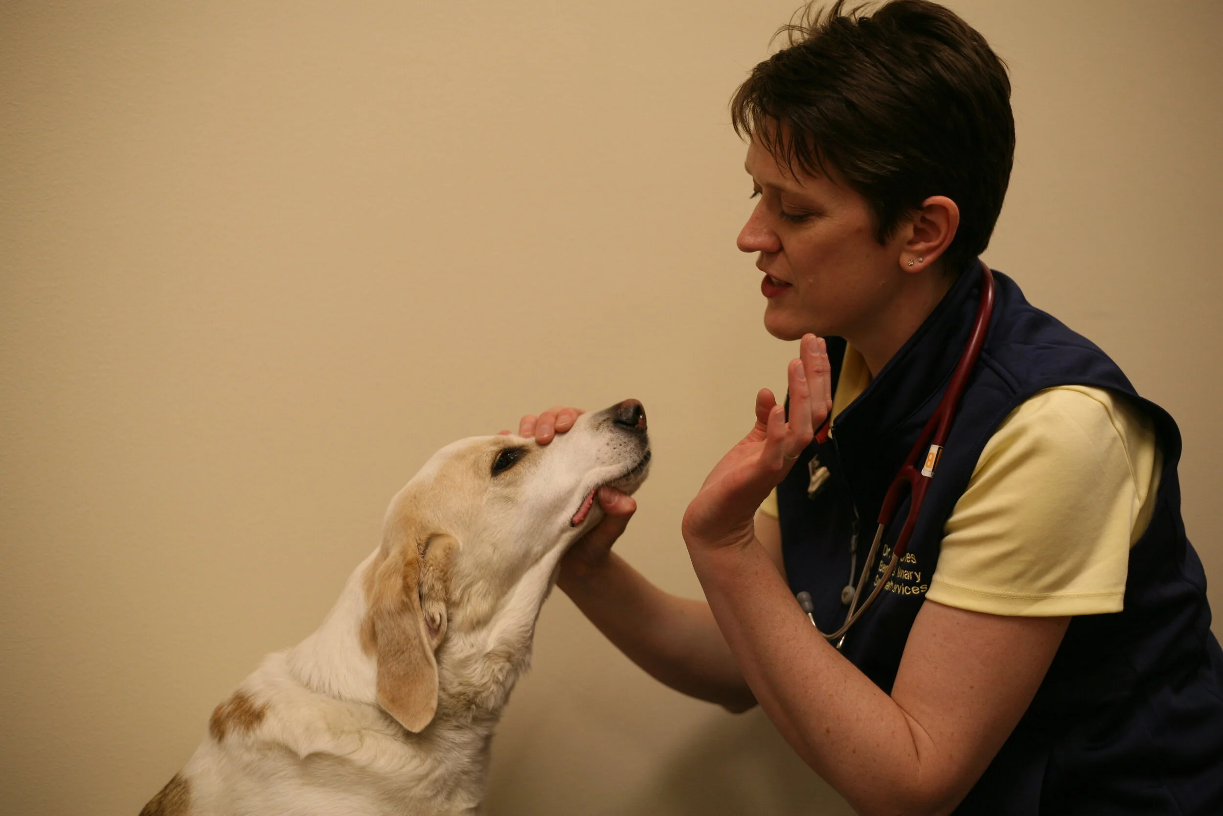
It’s an easy-to-do part of the neurologic exam but what does it mean? What do you do with an abnormal finding?
Can the neurologic examination predict disease?
Age, the Neurologic Examination and Prediction of Disease
Age isn't a disease, right? But, disease is associated with age. The older pet, is more likely to have structural disease (i.e. idiopathic epilepsy vs CNS neoplasia), compared to the younger pet. But, none of us want to diagnose a terminal disease in an older patient simply because the patient is older!
How can the Neurologic Examination Help Vets with Old Patients?
Can we look at two of the most commonly performed tests on the neurologic examination and determine the sensitivity and specificity for the detection of a forebrain lesion? Actually, yes we can. The menace response and paw replacement (previously called conscious proprioception) testing both assess the forebrain and are some of the most commonly performed parts of the neurologic examination. Here is what a recent group from Australia found:
Menace response
Sensitivity: 72%
Specificity: 47%
Odds ratio: 2.26
Proprioception
Sensitivity: 54%
Specificity: 72%
Odds ratio: 3.08
If age is then factored into the analysis, dogs greater than or equal to 6 years of age are more likely to have a forebrain disease detected if they have a menace or proprioceptive deficit.
As a "field" neurologist (without a pocket MRI...yet) this tells me that I should encourage diagnostic imaging in patients with menace deficits and proprioceptive deficits. The chances (or Odds) of a patient having underlying forebrain disease is higher if they have these deficits than if they don't. Seems intuitive, but proprioceptive testing isn't as sensitive as assessing the menace response so by all means don't forget to do that on an older patient! :)
Hope this little study was insightful for you too.
Chan MK, Jull P. Accuracy of selected neurological clinical tests in diagnosing MRI-detectable forebrain lesion in dogs [published online ahead of print, 2020 Jul 15]. Aust Vet J. 2020;10.
Keep those consults coming! I'm continuing to answer email consults in the evenings but do my best to be available during working hours should you have a questions and wish to call or text me. On site consultation is available Monday through Saturday at variable times throughout the week.
Have a good week!
Neuroanatomic Lesion Localization Practice Case
Lesion Localization and Case Building Practice
Lesion localization is one of those things that can be lost, if not practiced. Don't lose it! You're welcome! :)
Maria, is a 13 year old FS Lab
History: Presented to me with a 24 hour history of acute onset difficulty walking.
Neurologic examination:
Mentation: BAR
Cranial nerves: right head tilt, rotary nystagmus, remainder normal.
Gait: Moderate vestibular ataxia, falling right. No hypermetria or intention tremors noted.
Postural reactions: absent right thoracic and right pelvic limbs, normal other limbs
Spinal reflexes: Normal all limbs, normal c. trunci and perineal
Palpation: non painful, normal cervical ROM
You know what you've got to do now, right?
What is the Neuroanatomic Lesion Localization?
There are several ways to go through lesion localization.
OPTION 1:
I like to make lists. Start by listing all of the abnormalities and ALL possible locations that could result in an abnormal finding. For example:
1) Right head tilt - peripheral CN 8 (right), medulla (right), cerebellum (right or left)
2) Rotary nystagmus - same as above
3) Vestibular ataxia - same as above
4) Reduced paw replacement right side - right C6-T2, right C-C5, right medulla, right pons, right or left midbrain, left prosencephalon.
Now, we start to eliminate some things. How do to differentiate peripheral vs. brainstem vs cerebellar disease? Well, for starters any animal with cerebellar disease is expected to have hypermetria and/or intention tremors and Maria did not. We can cross out cerebellar disease. What else? Animals with brainstem disease should have a) change in mentation and/or b)ipsilateral paw replacement deficits and/or c) hemiparesis. Maria has paw replacement deficits ipsilateral to the head tilt so she most likely has brainstem disease.
OPTION 2:
The other way to work through this is to identify the cranial nerve affected on the exam (in this case, cranial nerve 8), identify the brainstem segment associated with this cranial nerve (in this case, medulla) and then ask yourself if you can identify a,b, or c from above. If not, it is peripheral and if so, it is brainstem.
Differential Diagnoses
Brain stem vestibular disease in an elderly dog without an important prior medical history would suggest the following differential diagnoses:
Degenerative: none
Anomalous: none
Metabolic: Hypothyroidism
Neoplastic/nutritional: Neoplasia of the brainstem
Infectious/inflammatory/idiopathic: meningoencephalitis (infectious or inflammatory)
Trauma: no supportive history
Vascular: Cerebrovascular accident (stroke)
Diagnostic plan
CBC, serum biochemistry, T4, blood pressure, urinalysis. (Screen for causes of stroke and hypothyroidism). Brain MRI +/- CSF tap.
Final diagnosis: Cerebrovascular disease! She was lucky. We didn't find any underlying cause of disease therefore this was considered idiopathic vascular disease. She showed gradual improvement with the addition of meclizine over 3-4 days with a residual head tilt after 7 days. The head tilt is expected to be permanent but isn't always the case.
How did you do? The neurologic examination, when done thoroughly, can be your best diagnostic tool for patients with neurologic disease! Keep practicing.
I hope you are well, and taking care of yourself. If you have a dog or cat with neurologic disease please reach out - I'd love to help.
Happy September 1st!
How the neurologic examination can help with seizure management
Movement
Are You Ready For a Tongue Twister?
What is the difference between paresis and ataxia and why should you care?
CPS, Postural Reactions, Paw Replacement...Oh My!
Performing CPs, or the paw replacement test, is so important for lesion localization, or to even identify if a patient has a neurologic problem. But it is difficult! This test causes residents to fret and frown because subtle differences in how you hold a patient, the flooring, and even environmental stimuli can affect how they place their limb. If you practice…it gets easier!


