neurologic exam

Neuroanatomic Lesion Localization for Busy Vets


Here is the case: A 5 year old cat that cannot blink one eye. What cranial nerve is affected?

To answer this question, of course you must do a cranial nerve exam. At its most basic level, it is a process of elimination. If you touch the medial and lateral canthus and the cat does not blink what cranial nerves are you testing? (CN 5 and CN 7)

How can you isolate these two nerves from each other to see which is the affected nerve? If you do corneal reflex you're testing CN 5 (sensory) and 6 (motor). Voila! So, if the cat does not blink when you touch the medial or lateral canthus, but DOES retract the globe when you do corneal reflex which nerve is affected? Think you know... scroll (or read!) to the bottom to see the answer. 

Need a little refresher on the cranial nerves and their jobs? The table below has all nerves, and their main jobs, for quick easy reference. 

Cranial nerveFunctionNeurologic examination CN I: OlfactorySmellWatch the dog sniff, ask about eating habits. * Difficult to objectively evaluateCN II: OpticSightMenace, PLR, Cotton ball test, trackingCN III: OculomotorSomatic: eye movement.
Innervates all extraocular muscles except lateral rectus and dorsal obliqueAutonomic: Parasympathetic function to the pupilPhysiologic nystagmus (medial movement), Strabismus (if present this indicates an abnormalities of CN)
 
PLRCN IV: TrochlearEye positionStrabismusCN V: Trigeminal

  • Ophthalmic

  • Maxillary

  • Mandibular

Sensory to the face, cornea
 
Close the jaw (muscles of mastication)Sensory: Corneal reflex, blink reflex, sensory assessment of the nares and lips
Motor: ability to CLOSE the jawCN VI: AbducentSomatic eye movementPhysiologic nystagmus (lateral movement), StrabismusCN VII: FacialSomatic: muscles of facial expression
Autonomic: parasympathetic innervation to the lacrimal gland, 3rd eyelid gland, palatine and nasal glands, taste rostral tongueMenace, blink reflex, lip and ear twitchCN VIII: vestibulocochlearSensory: balance and hearing
Innervates: vestibulospinal tracts, MLF (eye movement), reticular formation, cerebrum and cerebellumPhysiologic nystagmus, pathologic nystagmus,  positional strabismus, head position (tilt), ataxia, hearing test (BAER)CN IX: GlossopharyngealSensory to pharynx
Motor to pharynx
Autonomic: parasympathetic function – taste from the caudal tongueGag reflexCN X: VagusMotor to pharynx
Parasympathetic: taste to pharynx, larynx, heart rate, GI motility, otherGag reflex
CN XI: Spinal accessoryMotor: trapezius, sternocephalicus, brachiocephalicusPalpation of associated musclesCN XII: HypoglossalMotor: tongueMove tongue left and right, visually assess for symmetry and movement.


Answer: Cranial nerve 7 is affected. (5 is normal in corneal reflex therefore it is not the problem in the blink reflex either.)


Thanks for reading and have a great week!


 Do you have a case that is puzzling you? Please reach out - I'd love to help. Did you know I also do onsite or virtual private CE for hospitals? Reach out for more details, if you're interested.

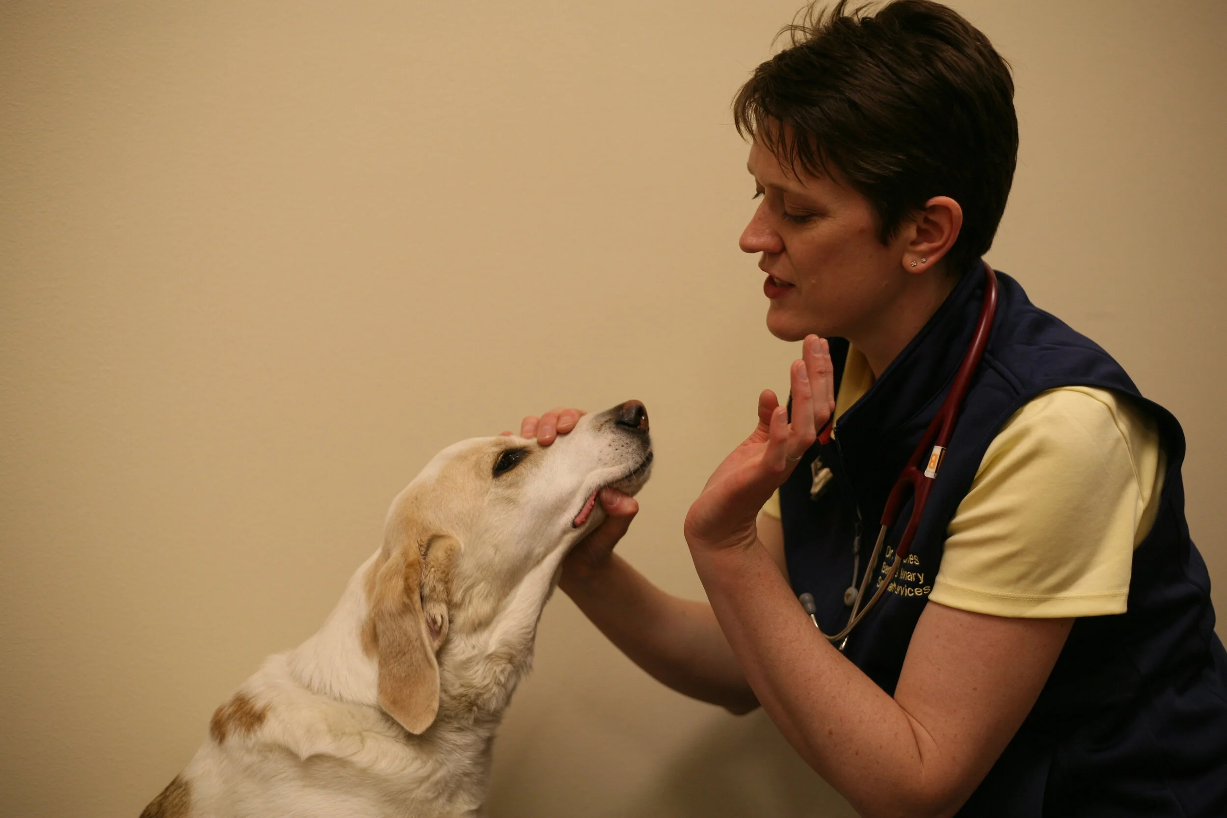
How reliable is the neurologic exam for patients with vestibular disease?

We (neurologists) like to think that the neurologic examination is the ultimate-be-all-end-all tool. But in dark corners, we talk about how incredibly hard it can be to do on patients with vestibular disease.
First, there are three parts that we need to consider for the lesion localization, correct?
1) Brainstem
2) Cerebellum
3) Peripheral CN 8
My rule of thumb is this: If the pet has ipsilateral hemiparesis/monoparesis, ipsilateral paw replacement deficits or decreased mentation (obtunded, stupor, coma) it is a brainstem lesion. If the pet has hypermetria, or intention tremors along with the vestibular signs, it is cerebellar in origin. Finally, in absence of those findings the lesion is localized peripherally.

An article out of Europe in 2019, dispelled our fears of the neurologic examination failing us and (thankfully) helped us sleep better at night when it was published that the neurologic examination correctly predicted if the vestibular signs were central (brainstem or cerebellum) or peripheral (cranial nerve 8) over 90% of the time.


Interestingly, central disease was more common in this study and, it was localized correctly MORE often than peripheral disease was localized correctly. In other words, dogs with central disease were more likely to be localized on the exam as having central disease compared to dogs with peripheral disease which were occasionally incorrectly localized with central disease.

A few more good reminders:

  • Nystagmus are not a localizing sign! (E.g. 8 dogs with peripheral and 5 dogs with central disease had horizontal nystagmus.)

  • The onset of disease does not predict it's lesion localization. (E.g. Acute and chronic onset of signs were not statistically different between the central and the peripheral groups.)

  • They had a lot of French Bulldogs in the study! Huh..I'm not sure I've noticed an over representation of French Bulldogs in my clinical work. It's good to learn something new everyday.

So, what does that mean for us?

It means if you do a thorough neurologic exam, you'll be correct about 90% of the time when you guide a client towards an MRI and spinal tap (for central disease) or treat for idiopathic or otitis (for peripheral disease). If you're unsure, err on the side of it being a central lesion and recommend a full work up. (Or contact me for a consult!) Oh, and 68% of dogs diagnosed with peripheral vestibular were idiopathic! Idiopathic disease means we have a lot more to learn...so let's get back to it!

(Bongartz U, et al. Vestibular Disease in dogs: association between neurological examination, MRI lesion localization and outcome. JSAP 2019).

Thanks for reading! This was an oldie, but a goodie and I hope you enjoyed revisiting it along with me. Please reach out if you have any questions. Have a great week

Is Age a Disease?

Age, The Neurologic Examination and Prediction of Disease


Age isn't a disease, right? No, it isn't but disease is associated with age. The older pet is more likely to have structural disease (i.e. neoplasia instead of idiopathic epilepsy), compared to the younger pet. That said, none of us want to diagnose a terminal disease in an older patient simply because the patient is older!

Can the Neurologic Examination Help Vets with Older Patients?


Let's look at the two most commonly performed parts of the neurologic examination and see how they related to disease. The menace response and paw replacement testing (previously called conscious proprioception) both assess the forebrain and are some of the most commonly performed parts of the neurologic examination. Here is what a recent group from Australia found in reference to finding evidence of forebrain disease on MRI:

Menace response
Sensitivity: 72%
Specificity: 47%
Odds ratio: 2.26

Proprioception
Sensitivity: 54%
Specificity: 72%
Odds ratio: 3.08

If age is then factored into the analysis, dogs greater than or equal to 6 years of age are more likely to have a forebrain disease detected by MRI if they have a menace or proprioceptive deficit.

As a "field" neurologist (without a pocket MRI...yet) this tells me that I should encourage diagnostic imaging in patients with menace deficits, and possibly for those with proprioceptive deficits depending on concurrent findings. The chances (or Odds) of a patient having underlying forebrain disease is higher if they have these deficits than if they don't. Seems intuitive, but proprioceptive testing isn't as sensitive as assessing the menace response.


Although this TidBit is a repeat from 2020, I liked this study and thought it was worth repeating.
Chan MK, Jull P. Accuracy of selected neurological clinical tests in diagnosing MRI-detectable forebrain lesion in dogs [published online ahead of print, 2020 Jul 15]. Aust Vet J. 2020;10.

Keep those consults coming! It has been a hectic week (for all of us, I think!) so remember to breathe, eat and hydrate. Have a great week!

How to Diagnose Idiopathic Epilepsy in the Exam Room

In 2015, the International Veterinary Epilepsy Task Force (IVETF) published a list of criteria to diagnose idiopathic epilepsy in dogs (not cats). The IVETF consisted of a group of veterinary neurologists, neuropathologists, and epileptologists. If you wish to see all of the resulting publications, just let me know! (They are available open access.)

The IVETF listed several ways to make a diagnosis of idiopathic epilepsy, starting with the lowest level of confidence (Tier I) and rising to the top with the most confidence in the diagnosis (Tier III). When using this information in your daily practice, consider writing "diagnosis: idiopathic epilepsy with Tier 1 confidence" in the medical record when a patient meets the criteria for a Tier I diagnosis of idiopathic epilepsy.

Please note, idiopathic epilepsy does not mean, any seizure disorder. Idiopathic epilepsy is a specific disease, that causes repeated seizures. ANY animal with repeated seizures can be diagnosed with epilepsy, but idiopathic epilepsy is ONE form of epilepsy. Make sense?


Tier I:

A dog must have:

  • A history of 2 or more seizures at least 24 hours apart

  • Have the age at onset between 6 months and 6 years

  • Demonstrate a normal inter-ictal physical and neurologic examination

  • A normal CBC, and serum biochemistry (the IVETF lists what they consider to be standard in a serum biochemistry analysis. Let me know if you want/need this list.)

  • A normal fasting bile acids and/or ammonia

  • A normal urinalysis

  • A familial history of IE is supportive, but not required


Tier II:

To diagnose idiopathic epilepsy with Tier II level confidence a dog must have:

  • All of Tier I plus...

  • A normal brain MRI

  • A normal CSF analysis

  • Normal fasting and post-prandial bile acids


Tier III:

  • All of Tier II plus...

  • Identification of ictal or inter-ictal EEG abnormalities suggestive of seizure disorders.

So, if a patient meets Tier I level confidence, when should MRI be performed?

  • Anytime a client wishes to confirm a diagnosis of intracranial disease. (This includes idiopathic epilepsy which is diagnosed by exclusion of other causes)

  • If a dog has a seizure onset of < 1 year or > 7 years of age (according to the IVETF)

  • If neurologic abnormalities reflective of the prosencephalon/forebrain are identified on the neurologic examination regardless of age, breed or a familial history of seizures.


In summary, it is always worthwhile to inform clients of the option of MRI when their dog (or cat) has seizures however, if the Tier I criteria are met and MRI has a high likelihood of normal results, it is very reasonable to skip this test and begin treatment for idiopathic epilepsy.

I try to remember that MRI abnormalities were identified in 22% of dogs with a normal neurologic exam and 90% of dogs with an abnormal neurologic examination in one study.


What about CSF analysis, you ask? I'm glad you asked! Some dogs with meningitis will have a normal MRI, therefore an MRI PLUS CSF tap is often my recommendation to ensure we don't overlook those patients inappropriately by just performing an MRI.

What about cats? The IVETF recommendations do not specifically apply to cats however many neurologists, including me, extrapolate this information to cats in practice. My fingers and toes are crossed for science driven cat-specific recommendations in the near future.


Have a great week! As always stay safe, and let me know how I can help you, help your patients, with neurologic disease.

Neurologic Cat?

Where is the Lesion?

History: Sarah is a 4 year old, indoor only FS DSH. She was obtained as a kitten and had been normal, per clients, her entire life thus far. She presented to me for acute onset (same day) weakness. No known trauma, toxin ingestion or medication exposure.

Physical examination: No abnormalities, normal TPR.

Neurologic examination:
•Mentation: BAR
•Gait: Ambulatory severe left hemiparesis with proprioceptive ataxia in all four limbs
•Cranial nerves: Normal
•Postural reactions: absent left thoracic and pelvic limbs only
•Reflexes: reduced withdrawal left thoracic limb, normal all other limbs. Cutaneous trunci absent left, normal right side.
•Palpation: Non-painful

Lesion localization? I do not see evidence of intracranial disease so automatically I localize caudal to C1. Both a front and rear leg are affect, so again, we can isolate cranial to T2. So...right away you can think left C1-C5 or C6-T2 myelopathy. Which is it? Well, the reflexes were reduced to the left thoracic limb, and the reflex arc is C6-T2, so our localization must involve C6-T2.
Up is differential diagnoses building. Here is what I came up with:
D: none, this is acute
A: None, the cat is too old for congenital disease onset signs
M: none
N: Lymphoma is possible however it is rarely acute in nature
I: Meningomyelitis (infectious or inflammatory) is possible
T: No known trauma, but cannot rule it out
V: Fibrocartilagenous embolism is highly likely due to the acute onset of signs.

Did you think of something else that I missed?
Unabashed plug for the CE August 2nd here - we will look more specifically at this case and do lesion localization in greater detail. Please consider joining us from 7-8PM. Details can be found at my website.

Diagnosis: I debated about giving this away before the talk and I have decided NOT to tell you the final diagnosis this week. I will share it on next week's TidBit Tuesday (August 3rd) so stay tuned!

Hopefully you enjoyed this case review this week. Do you have a suggestion for a TidBit Tuesday topic? If so - please send me an email. I would prefer to write about something you want to read. :)

I hope you have a great week!

The Neurology Checklist

The Checklist

Odds are high that you have probably heard about surgical or operative checklists and, perhaps, some of you employ them in your clinic. This week I wanted to revisit this idea because a recent article reminded me of the benefit of checklist so I thought I'd share it with you!


How Does it Work?

You and your team devise a list of important things to remember for a given procedure and create a checklist. This list then hangs in your OR, or outside of a lab, or wherever it will be used. Ten years ago I read a book called The Checklist Manifesto (https://www.amazon.com/Checklist-Manifesto-How-Things-Right/dp/0312430000). One of the simplest ways to save a life is to know the name of your anesthetist and lead surgeon. Yep, that's it. Instead of bumbling around and saying "hey you...anesthesia..." you can address them by their name and shorten communication time in a crisis. Perhaps you work very closely with your staff everyday and it seems inconceivable that you wouldn't know someone's name! However for new staff members, or in large clinics, this may not be the case.


What is the evidence?

A study published in the Journal of Veterinary Surgery this month identified that surgical times were significantly shorter (p= .005) , antibiotic administration (when appropriate) was given as intended more often (p=.01), and fewer dogs had an unplanned return to the OR (p=.006) when a perioperative checklist was employed. (Reference below.) It takes less than 1 minute to run through the checklist and it may change the course of you or your patient's day!


What is the Take Home Message?

If you don't already have one, consider making a checklist for any procedure in which patient care could be compromised if something was forgotten. This doesn't need to apply ONLY to surgical situations! I make an effort to know the name of my assistant during each and every neurologic examination for this exact reason. I don't expect a crisis during a neurologic examination but everyone feels more committed to the team when we ARE a team! (And I like to be friendly, too!) I also had a checklist hanging in the neurology room when I was at UW and at VCA Aurora to remind trainees of the parts of the neurologic examination.

Neuro Checklist:
Mentation
Cranial nerves
Gait assessment
Postural reactions
Reflexes
Palpation
Ask about a seizure history.

Having this list in your work space ensures that you don't accidentally forget to perform part of the neurologic examination and yet, it isn't so overwhelming that you hesitate to read it. (A checklist MUST be short or people hate to do them!)


I hope you enjoyed this not-fully-totally-neurology-related TidBit Tuesday. Please let me know if you have any comments, questions, or requests for topics. Have a great week and keep those consults coming!

Reference: Thieman Mankin, KM, Jeffery ND, Kerwin SC. The impact of a surgical checklist on surgical outcomes in an academic institution. https://doi.org/10.1111/vsu.13629


Are we any good at a neurologic exam when pets are vestibular?

How reliable is the neurologic exam for patients with vestibular disease?

We (neurologists) like to think that the neurologic examination is the ultimate-be-all-end-all tool. But in dark corners, we talk about how incredibly hard it can be to do on patients with vestibular disease.
First, there are three parts that we need to consider for the lesion localization, correct?
1) Brainstem
2) Cerebellum
3) Peripheral CN 8
My rule of thumb is this: If the pet has ipsilateral hemiparesis/monoparesis, ipsilateral paw replacement deficits or decreased mentation (obtunded, stupor, coma) it is a brainstem lesion. If the pet has hypermetria, or intention tremors along with the vestibular signs, it is cerebellar in origin. Finally, in absence of those findings the lesion is localized peripherally.

An article out of Europe, dispelled our fears of the neurologic examination failing us and (thankfully) helped us sleep better at night when it was published that the neurologic examination correctly predicted if the vestibular signs were central (brainstem or cerebellum) or peripheral (cranial nerve 8) over 90% of the time.


Interestingly, central disease was more common in this study and, it was localized correctly MORE often than peripheral disease was localized correctly. In other words, dogs with central disease were more likely to be localized on the exam as having central disease compared to dogs with peripheral disease which were occasionally incorrectly localized with central disease.

A few more good reminders:

  • Nystagmus are not a localizing sign! (E.g. 8 dogs with peripheral and 5 dogs with central disease had horizontal nystagmus.)

  • The onset of disease does not predict it's lesion localization. (E.g. Acute and chronic onset of signs were not statistically different between the central and the peripheral groups.)

  • They had a lot of French Bulldogs in the study! Huh..I'm not sure I've noticed an over representation of French Bulldogs in my clinical work. It's good to learn something new everyday.

So, what does that mean for us?

It means if you do a thorough neurologic exam, you'll be correct about 90% of the time when you guide a client towards an MRI and spinal tap (for central disease) or treat for idiopathic or otitis (for peripheral disease). If you're unsure, err on the side of it being a central lesion and recommend a full work up. (Or contact me for a consult!) Oh, and 68% of dogs diagnosed with peripheral vestibular were idiopathic! Idiopathic disease means we have a lot more to learn...so let's get back to it!

(Bongartz U, et al. Vestibular Disease in dogs: association between neurological examination, MRI lesion localization and outcome. JSAP 2019).

My current work days are...well, all of them except Sundays. I'll post on FB or my website if I'm closed on a random day so feel free to check those spots if you're not sure. Otherwise, feel free to call, email or hop online to schedule a telephone, live or video consultation with me. Remember, all live consults are still curbside!

Can the neurologic examination predict disease?

Age, the Neurologic Examination and Prediction of Disease


Age isn't a disease, right? But, disease is associated with age. The older pet, is more likely to have structural disease (i.e. idiopathic epilepsy vs CNS neoplasia), compared to the younger pet. But, none of us want to diagnose a terminal disease in an older patient simply because the patient is older!

How can the Neurologic Examination Help Vets with Old Patients?


Can we look at two of the most commonly performed tests on the neurologic examination and determine the sensitivity and specificity for the detection of a forebrain lesion? Actually, yes we can.  The menace response and paw replacement (previously called conscious proprioception) testing both assess the forebrain and are some of the most commonly performed parts of the neurologic examination. Here is what a recent group from Australia found:

Menace response
Sensitivity: 72%
Specificity: 47%
Odds ratio:  2.26

Proprioception
Sensitivity: 54%
Specificity: 72%
Odds ratio: 3.08
If age is then factored into the analysis, dogs greater than or equal to 6 years of age are more likely to have a forebrain disease detected if they have a menace or proprioceptive deficit. 

As a "field" neurologist (without a pocket MRI...yet) this tells me that I should encourage diagnostic imaging in patients with menace deficits and proprioceptive deficits. The chances (or Odds) of a patient having underlying forebrain disease is higher if they have these deficits than if they don't. Seems intuitive, but proprioceptive testing isn't as sensitive as assessing the menace response so by all means don't forget to do that on an older patient! :)

Hope this little study was insightful for you too.
Chan MK, Jull P. Accuracy of selected neurological clinical tests in diagnosing MRI-detectable forebrain lesion in dogs [published online ahead of print, 2020 Jul 15]. Aust Vet J. 2020;10.

Keep those consults coming! I'm continuing to answer email consults in the evenings but do my best to be available during working hours should you have a questions and wish to call or text me. On site consultation is available Monday through Saturday at variable times throughout the week.

Have a good week! 

Neuroanatomic Lesion Localization Practice Case

Lesion Localization and Case Building Practice

Lesion localization is one of those things that can be lost, if not practiced. Don't lose it! You're welcome! :)


Maria, is a 13 year old FS Lab
History: Presented to me with a 24 hour history of acute onset difficulty walking. 

Neurologic examination:
Mentation: BAR
Cranial nerves: right head tilt, rotary nystagmus, remainder normal.
Gait: Moderate vestibular ataxia, falling right. No hypermetria or intention tremors noted. 
Postural reactions: absent right thoracic and right pelvic limbs, normal other limbs
Spinal reflexes: Normal all limbs, normal c. trunci and perineal
Palpation: non painful, normal cervical ROM

You know what you've got to do now, right?

What is the Neuroanatomic Lesion Localization?

There are several ways to go through lesion localization.

OPTION 1:
I like to make lists. Start by listing all of the abnormalities and ALL possible locations that could result in an abnormal finding. For example:
1) Right head tilt - peripheral CN 8 (right), medulla (right), cerebellum (right or left)
2) Rotary nystagmus - same as above
3) Vestibular ataxia - same as above
4) Reduced paw replacement right side - right C6-T2, right C-C5, right medulla, right pons, right or left midbrain, left prosencephalon.

Now, we start to eliminate some things. How do to differentiate peripheral vs. brainstem vs cerebellar disease? Well, for starters any animal with cerebellar disease is expected to have hypermetria and/or intention tremors and Maria did not. We can cross out cerebellar disease. What else? Animals with brainstem disease should have a) change in mentation and/or b)ipsilateral  paw replacement deficits and/or c) hemiparesis. Maria has paw replacement deficits ipsilateral to the head tilt so she most likely has brainstem disease. 

OPTION 2:
The other way to work through this is to identify the cranial nerve affected on the exam (in this case, cranial nerve 8), identify the brainstem segment associated with this cranial nerve (in this case, medulla) and then ask yourself if you can identify a,b, or c from above. If not, it is peripheral and if so, it is brainstem. 

Differential Diagnoses


Brain stem vestibular disease in an elderly dog without an important prior medical history would suggest the following differential diagnoses:
Degenerative: none
Anomalous: none
Metabolic: Hypothyroidism
Neoplastic/nutritional: Neoplasia of the brainstem
Infectious/inflammatory/idiopathic: meningoencephalitis (infectious or inflammatory)
Trauma: no supportive history
Vascular: Cerebrovascular accident (stroke)

Diagnostic plan

CBC, serum biochemistry, T4, blood pressure, urinalysis. (Screen for causes of stroke and hypothyroidism). Brain MRI +/- CSF tap. 

Final diagnosis: Cerebrovascular disease! She was lucky. We didn't find any underlying cause of disease therefore this was considered idiopathic vascular disease. She showed gradual improvement with the addition of meclizine over 3-4 days with a residual head tilt after 7 days. The head tilt is expected to be permanent but isn't always the case. 

How did you do? The neurologic examination, when done thoroughly, can be your best diagnostic tool for patients with neurologic disease! Keep practicing. 

I hope you are well, and taking care of yourself. If you have a dog or cat with neurologic disease please reach out - I'd love to help. 

Happy September 1st!