What Do You Do With a Confused Cat?

What do you do with a confused cat?

Sigalment: 12 year old FS Domestic short-haired cat
History: 2-3 week history of seeming "confused", and a recent onset of circling right more than left. These very astute owners observed the cat seemed to walk into a room, forget why it entered, stand there, and then leave again. Additionally, she wasn't as bright as is typical for this indoor-outdoor Wisconsin cat. Birds would land on the feeder outside of the window and she appeared uninterested. 
Physical examination: Normal TPR, normal exam. This was a remarkably fit cat for her age!
Neurologic examination:
Mentation: Obtunded
Cranial nerves: absent left menace response, normal PLR and normal menace response on the right. The cat was less responsive to facial stimulation on the left lip but did respond to hemostat pinch. All other cranial nerve exam findings were within normal limits. 
Gait: Ambulatory with mild proprioceptive ataxia and occasional circling right
Postural reactions: absent tactile placing left pelvic limb, intermittent on left thoracic limb, normal on right thoracic and pelvic limbs. 
Spinal reflexes: normal all limbs, and c. trunci

Okay..take a moment and put all of that together in your mind. I'll take this moment to remind you that my next Webinar is on Wednesday July 15th (TOMORROW) and we'll be talking about lesion localization. If you're interested in joining me to practice on cases just like this please go to https://barnesveterinaryservices.com/ce-opportunities and sign up!

Back to this case. How did you do? Here is how I would talk myself through this one...
1. The cat has a change in mentation..the problem MUST be intracranial.
2. There is a menace deficit - the affected parts could be cranial nerve 2, forebrain or cranial nerve 7. 

  • 2a. PLR is normal (this is cranial nerve 2, midbrain and cranial nerve 3) so we can eliminate cranial nerve 2 as a possible cause of the menace deficit. 

  • 2b. The cat did not have any noted change to motor of the face (run by cranial nerve 7), so we can eliminate cranial nerve 7 as a cause for the menace deficit. 

  • 2c. Finally, one must remember that the menace response crosses and enters the opposite side forebrain so, since we have narrowed the problem down to the forebrain we then must comment that it is the RIGHT forebrain we are concerned about

3. Circling is either a vestibular or forebrain sign. This cat did not have evidence of vestibular disease so we could consider this another forebrain sign. Animals circle towards their lesion, thus supporting a RIGHT forebrain lesion. 

So far, so good?

4. Tactile placing goes from the toes, up the ipsilateral spinal cord, brain stem (crossing at the midbrain) and enters the opposite side forebrain. Voila! We don't have any evidence of spinal cord disease, or brain stem disease in this cat (no cranial nerve deficits) so this also suggests a RIGHT forebrain lesion. 

What about that sensory deficit on the cranial nerve exam? This actually suggests a proprioceptive change to the face! This finding might be the only finding we didn't routinely teach in veterinary school because it's an oddity. But, it is a crossing tract and reflects forebrain disease. 

So, what lesion localization did you come up with? Scroll to the bottom to see if you are correct.

Diagnosis
We diagnosed a brain tumor on MRI (see image above) and took this kitty to surgery. The mass was removed entirely, and was eventually diagnosed as a meningioma. Meningiomas are locally invasive, rarely spreading, intracranial tumors in cats. With complete surgical resection no additional treatment is needed! The cat went back to her normal personality, stopped circling, and regained vision in the left eye post operative (by the 1 month recheck...not immediately). Pretty cool stuff, huh!?

I hope you have a safe and fun week. Keep those consults coming and I hope to see you at the Webinar tomorrow!

Answer: RIGHT forebrain. 

What drug, route and dose should you use for at home acute seizure management?

I hope you had a wonderful and safe 1st and 4th of July (my American and Canadian friends)! Enjoy this week's TidBit Tuesday...

What drug, route and dose do you use for at home acute seizure management?

For most of us, benzodiazepine drugs (diazepam, midazolam, lorazepam) are our first choice drugs for acute seizure cessation.

Not all benzodiazepine drugs are equal!
What routes can you give the drugs? What routes shouldn’t you give the drugs?


Benzodiazepine drugs were introduced in the 1960s for human status epilepticus. A 2015 human meta-analysis identified that benzodiazepines are the “best” first line IV drugs. To date, there have not been any veterinary studies identifying which drug is ‘best’ for acute seizure management. We’ve just always used benzodiazepines so we continue to do so. Furthermore, limited data regarding efficacy is available for veterinary patients. So, what do we know?? The table below outlines the approved routes for each of the common benzodiazepine drugs. This should be printable. 


DiazepamSafe for dogs ☒Safe for cats ☒            Okay if given:IV ☒IM ☐ Not recommended!Rectal ☒  Preferred drug!Intranasal ☒MidazolamSafe for dogs ☒Safe for cats ☒            Okay if given:IV ☒IM ☒Rectal ☐ (nope).Intranasal ☒  Preferred drug!LorazepamSafe for dogs ☒Safe for cats ☒            Okay if given:IV ☒IM ☐ No dataRectal ☐ Nope, not absorbedIntranasal ☒


In 2017 a randomized veterinary clinical trial determined that intranasal midazolam was superior to rectal diazepam for controlling seizures in some dogs. As such, intranasal midazolam (0.2 mg/kg intranasal) is currently my treatment recommendation for at home anti-convulsant care. Guess what? It works great in hospital too when you cannot access a vein fast enough. 

My current recommendations for at home care are:
1) Intranasal midazolam at 0.2 mg/kg IN up to 3x in 24 hours. Use a nasal atomizer - it's a game changer. 
2) Rectal diazepam at 1 mg/kg PR up to 3x in 24 hours. Make sure to prescribe the BOTTLE, this drug is light sensitive so dosing it in a syringe makes it useful for up to 30 minutes and then it may loose potency. 

Other choices, such as rectal or IV levetiracetam, have been evaluated for acute seizure management too. We have more data about benzodiazepine drugs, and I'm more accustomed to using them (over the past 20 years!) therefore they are still my preference. I'll keep you posted if data surfaces suggesting we should change to using levetiracetam, or another drug, instead of the benzodiazepine class. 

Stay safe and keep those consults coming! For July my days of operation should remain Monday-Saturday. The schedule will change at the end of August once we sort out what kind of schooling we will be doing for the kiddos - I will keep you posted or you can always check the website or Facebook.  Thanks for reading!

Portosystemic Shunts and Their Effect on the Nervous System

Portosystemic Shunts and Their Effect on the Nervous System


Portosystemic shunts (PSS) are common in small breed dogs and surgical correction is commonly recommended. However, there is concern about neurologic signs peri-operative for these patients. Some develop seizures, mentation changes, and other neurologic signs before shunt attenuation while others do not show signs until after shunt attenuation. Sadly, the presence of neurologic signs is a poor prognostic indicator according to many studies. One recent study looked for prognostic indicators in dogs that develop seizures post attenuation (PAS) within 7 days of surgery.  I hope you'll find the study as interesting as I did!

Study Design

This was a retrospective study including 93 dogs from 14 different institutions. 53% of dogs received prophylactic levetiracetam. There were lots of different small breeds with a median age of 34 months (range 5-124 months). 

Results

  • 17.3% of the dogs had experienced at least 1 seizure prior to surgery.

  • Interestingly, 78% of the dogs had exhibited neurologic abnormalities including those with seizures.

  • Only 30% of dogs with PAS survived to 30 days. Yikes.

  • More often those with focal seizures survived, compared to dogs with generalized seizures.

  • Sadly, the most common reason for euthanasia following the shunt was the presence of uncontrolled seizures. 

Key Points

According to their multivariate analysis the only two factors associated with short term survival were 1) having a history of seizures PRIOR to shunt correction (p=.004) and 2) the development of focal PAS (p=.0003). That's it! 

So, what do we tell our clients? Well, for starters..they need to know that these pets are are risk of developing seizures even if they DON'T have surgery. But, according to this study if they do elect surgery, the development of generalized seizures is a poor prognostic indicator. 

This article was packed with other data so if you want to read the entire thing please reach out and let me know. 

Reference:
Mullins RA, Sanchez Villamil C, Selmic LE, et al. Prognostic factors for short term survival of dogs that experience post attenuation seizures after surgical correction of single congenital extrahepatic portosystemic shunts: 93 cases (2005-2018) Vet Surg.2020; 49:958-970.


That's it for now! I'm maintaining curbside service for the summer and am starting to widen my travel radius again. Please reach out if I can asist with a case. Stay safe, stay healthy!

Help! My patient sustained head trauma!

Help! My Patient Sustained Head Trauma!


Traumatic brain injury (TBI) in dogs and cats is a dynamic process. The outcome depends not only on the severity of the primary injury, but also on the resulting secondary effects. Primary injury is defined as injury that occurs at the moment of impact and results in physical disruption of tissues (fracture, etc.).  Secondary injury is defined as the physiologic alterations that occur hours to days after the primary injury. Medical therapy is aimed at affecting secondary injury. Animals with TBI frequently have serious injuries elsewhere and shock, hemorrhage, airway obstruction, pneumothorax, and traumatic cardiac arrhythmias should be detected and treated timely. 
 
Pathophysiology
Following TBI local vascular disruption can cause hemorrhage, which results in deposition of iron, free radical formation and mass effect. The effects on the vascular system contribute to vasogenic edema. Cytotoxic edema is the other type of edema that can occur in head trauma. Cytotoxic edema forms when excessive neurotransmitters are released by surrounding cells, which cause overstimulation of neurotransmitter-dependent channels, causing excessive intracellular accumulation of sodium and calcium. Increased intracellular sodium results in an osmotic draw of water into the cell, causing swelling and possibly apoptosis.

Cerebral Perfusion and the Cushing's Reflex
Cerebral perfusion is the driving force behind cerebral blood flow and cerebral blood flow is reduced in the first 24 hours following traumatic brain injury. Reductions in cerebral blood flow can result in poor delivery of oxygen and metabolic substrates and inadequate removal of waste and CO2. CO2 is a potent vasodilator, therefore excessive local CO2 may result in local (or global if severe enough) vasodilation. Maintaining cerebral perfusion, and therefore cerebral blood flow is critical in the head injured patient. There are several equations that are commonly used in TBI and they include:

CPP = MAP – ICP1
CBF = CPP/CVR

CPP = cerebral perfusion pressureCBF = cerebral blood flowMAP = mean arterial blood pressureCVR = cerebral vascular resistanceICP = intracranial pressure


Animals with TBI may develop focal or global increases in CO2 as cerebral blood flow decreases, which result in vasodilation and therefore causes worsening CBF. Following TBI, the brain’s intrinsic ability to manage cerebral blood flow and perfusion pressure becomes altered. Vasomotor centers in the brain may detect increased CO2 and attempt to vasoconstrict through increase in sympathetic function. Systemic hypertension then occurs, which is noted clinically as an increase in MAP. This increase is detected by baroreceptors and a reflexive bradycardia ensues. Animals with decreased mentation, hypertension and bradycardia are at increased risk of having or eminently developing increased ICP. This triad of abnormalities (bradycardia, hypertension and altered mentation) in a post-traumatic patient is called the Cushing’s reflex. The end result of excessive increased ICP is brain herniation.

Management of the Head Trauma Patient
1) Get oxygen to the brain:  A clear airway must be established, oxygen and CO2 status should be determined. If oxygen supplementation is required, an oxygen cage or mask may be used. Remember to avoid nasal canula as sneezing can increase ICP. Also, oxygen supplementation will correct hypoxemia, but will not prevent hypercarbia in a hypoventilating animal. Tracheal intubation and mechanical ventilation is indicated in animals that are apneic or hypoventilating.
.
2) Monitor blood pressure and heart rate: Head-injured patients require maintenance of systemic and cerebral hemodynamics. The two most important goals are preservation of CPP and maintenance of systemic oxygen availability. Ideally mean arterial blood should be constantly monitored in these patients. Hemodynamic goals include a MAP>80-90 mm Hg and <115-120 mm Hg. Fluid restriction is NOT encouraged in the post-traumatic patient because of the risks to systemic health and the minimal benefit to reducing cerebral edema. Colloidal support may improve blood flow (and perfusion) however, the 2007 SAFE study in human TBI demonstrated an increased mortality in patients receiving albumin compared to those receiving saline rehydration. It remains unknown if all colloidal support would have the same outcome.3 
3) Manage Increasing intracranial pressure: Mannitol (1 gm/kg slow IV over 20-30 minutes) has become a cornerstone in the management of increased ICP. There are several proposed mechanisms of action by which mannitol decreases ICP. 1) It expands circulating volume, decreases blood viscosity and therefore increases cerebral blood flow and cerebral oxygen delivery. 2) Reduction of CSF production 3) free radical scavenger   4) Delayed effect:  osmotic action: transfer of extravascular edema fluid (in neurons) into the intravascular space: occurs 15-30 minutes after administration when gradients are established between plasma and cells. The effects of mannitol persist for a variable period ranging from 1 to 3 hours depending on clinical conditions. Mannitol can be administered as repeated boluses, or continuous infusion.  Due to a rebound effect, risk of hypernatremia and hyperosmolarity, mannitol should not be administered more often than 3 times over a 24 hour period. Dangers of repeated dosage are related to effects on blood volume and electrolytes rather than specific toxicity and the patient's blood volume status should be closely observed. Hypertonic saline may also be used instead of mannitol and is considered equivalent by most neurologists for most patients. Hypertonic saline (4 mL/kg of 7.5% or 5.4 mL/kg of 3%) has the added advantage of rapidly restoring volume by causing an osmotic shift from extracellular to intravascular space. Due to the high sodium level, salt toxicity is possible therefore; serum sodium levels should be frequently monitored.  The exception may be patients that are volume depleted; these patients may benefit from hypertonic saline more than mannitol. For a favorable prognosis, a response to medical therapy should be seen within 4 to 6 hours following commencement of treatment. An animal should be assessed every 30 minutes until stabilized.
Glucocorticoids are contraindicated in TBI due to the exacerbation of hyperglycemia.  The majority of available evidence indicate that glucocorticoids do not lower ICP, or improve outcome in severely head-injured patients.

Monitoring of TBI patients
1) respiratory status, pupil size (and PLR) and level of mentation. In a very simplistic sense, inspiration and expiration are initiated in the medulla, and the rate and pattern is driven from the midbrain/pontine centers. Feedback loops, largely responsive to blood levels of CO and oxygen are present in the prosencephalon as well. Damage to specific areas of the brain will result in specific respiratory patterns.
2) Monitoring pupil size can help with formulating a prognosis (see below) and aid in initial lesion localization.  Sympathetic function to the eye originates in the thalamus, descends through all parts of the brainstem and exits the spinal cord T1-T3. Damage to this tract will result in an inability to dilate the eye, or miosis. This most often occurs with damage to the thalamus (prosencephalon) or medulla. Parasympathetic innervation to the eye starts in the midbrain and goes to the eye by traveling along with the somatic fibers of cranial nerve 3. Damage to the midbrain would result in an inability to constrict the eye, or a dilated fixed pupil. If both the sympathetic and parasympathetic fibers are damaged the eye will appear fixed and midrange.
3) Monitor mentation changes are described as obtunded, stupor or coma. Obtunded animals have diminished responsiveness to the environment but will respond to tactile, visual or verbal stimuli. Animals with stupor will only respond to firm tactile stimuli (not visual or verbal) and animals in a coma will not respond to sharp tactile stimuli such as pinching with a hemostat. Body posture can also help one lesion localize the problem. Decerebrate rigidity occurs with damage to the descending corticospinal tracts, usually at the level of the midbrain. Animals have increased extensor tone to all limbs and severely decreased mentation, usually coma. This indicates a poor prognosis. Decerebellate posture occurs with damage to the cerebellar peduncles and typically has a fair to good prognosis. Patients demonstrate forelimb extension, variable pelvic limb positioning and normal mental awareness.
 
Prognosis
Both the modified Glasgow coma scale (MGCS) and animal trauma triage (ATT) score have been used to provide estimated, objective assessments for prognostication in animals. For the MGCS, motor activity, brainstem function and level of consciousness are scored  and then compared to a graph to determine the relative survival probability.5  For the MGCS, the LOWER the number, the LOWER the probability of survival. Remember to perform 2-3 GCS before considering the number reliable if the animal is in the immediate post-traumatic period. This scale is useful for adjusting the prognosis on a daily basis, as the patient progresses through treatment.
Unlike the MGCS, the animal trauma triage score accounts for more than just intracranial trauma. Scores are summed for each of the following six categories: perfusion, cardiac, respiratory, eye/muscle/integument, skeletal and neurological. Scores are 0-3 for each category resulting in a maximum score of 18. The LOWER the number the HIGHER the probability of survival. 6 For each increase in 1 point, there is a 2-2.6x decrease in survival.7 One recent study compared the two scoring systems for animals with head trauma and identified the ATT as a more predictive and reliable scoring system.7
 
References:
1.          Kuo KW, Bacek LM, Taylor AR. Head Trauma. Vet Clin NA Small Anim Pract. 2018;48(1):111-128.
2.          Van Beek J, Mushkudiani N, Steyerber E, et al. Prognostic value of admission laboratory parameters in traumatic brain injury: Results from the IMPACT study. J Neurotrauma. 2007;24(2):315-328.
3.          SAFE study I, Australian and New Zealand Intensive Care Society Clinical Trials G, Australian Red Cross Blood S, George Institute for International H, Myburg J, Cooper D. Saline or albumin for fluid resuscitation in patients with traumatic brain injury. N Engl J Med. 2007;357(9):874-884.
4.          Hayes GM. Severe seizures associated with traumatic brain injury managed by controlled hypothermia, pharmacologic coma, and mechanical ventilation in a dog: Case report. J Vet Emerg Crit Care. 2009;19(6):629-634.
5.          Platt SR, Radaelli ST, McDonnell JJ. The prognostic value of the modified Glasgow Coma scale in head trauma in dogs. J Vet Intern Med. 2001;15(6):581-584.
6.          Rockar RA, Drobatz KS, Shofer FS. Development of a scoring system for the veterinary trauma patient. J Vet Emerg Crit Care. 1994;4(2):77-83.
7.          Ash K, Hayes GM, Goggs R, Sumner JP. Performance evaluation and validation of the animal trauma triage score and modified Glasgow Coma Scale with suggested category adjustment in dogs: A VetCOT registry study. J Vet Emerg Crit Care. 2018;28(3):192-200.


That's it for now! I'm maintaining curbside servcie for the summer and am starting to widen my travel radius again. Please reach out if I can assit with a case. Stay safe, stay healthy!

Prevalence of Idiopathic Epilepsy in Dogs

Idiopathic Epilepsy Update!


A recent article out of the Vet Record by Dr. Rachel Hall and colleagues outlines the prevalence of idiopathic epilepsy and structural epilepsy in dogs.* I found this a very interesting read, packed with useful information so I thought I'd pass along a bit (get it?) of it to you!

Study Design and Points Worth Noting

  • This is a retrospective study based out of the UK. 

  • 900 cases with MRI, a neurologic examination and medical record history were included. (wow!)

  • Structural epilepsy is defined as a seizure disorder secondary to an identifiable structural cause. Examples include neoplasia, meningoencephalitis, hydrocephalus, etc.

  • Idiopathic epilepsy is defined as the lack of identification of a structural abnormality in a pet with 2 or more discrete seizures. 

  • Small (< 10 kg), medium (10-20 kg) and large breed (>20 kg) dogs were represented in approximately the same percentage in this study.

Results of Interest (there are a lot of interesting results in this study!)

  • About 50% of the dogs were between 6 months and 6 years old, and 50% were > 6 years old. 

  • About half of the dogs had structural epilepsy based on abnormal MRI findings

  • The other half of dogs had no significant findings on MRI and the majority were classified as having idiopathic epilepsy. (The others had toxin and metabolic disease diagnosed).

    • Prevalence of idiopathic epilepsy in dogs in the UK? 50%!


Okay, fine (you might think) how does knowing the prevalence of idiopathic epilepsy in dogs in the UK help me?

I'm glad you asked...

  • Idiopathic epilepsy was the leading diagnosis for dogs between 7 months and 6 years old. Inflammatory brain disease was second.

    • Take away point? Meningoencephalitis is NOT rare! Oh, and idiopathic epilepsy is most common in the group of dogs we thought it would be most common.  

  • Idiopathic epilepsy was also the leading single diagnosis (34%) for dogs over 6 years of age. HOWEVER when they combined all types of neoplasia together into one group they found 43% of dogs over 6 years of age had neoplasia making it the leading singe diagnosis in this group.

    • Take away point? Read that sentence above again! I considered making this TidBit Tuesday a one line update because it's so critical to make sure we don't forget that "old" dogs can actually have idiopathic epilepsy!

    • Also, dogs over 6 years of age had structural epilepsy more often than idiopathic epilepsy if all causes for structural epilepsy were combined. (Not surprising, I know.)

What do you do with this information?

Do a neuro exam on every patient with a seizure history!

  • If the exam is NORMAL, include idiopathic epilepsy on the differential diagnoses list, regardless of age. 

  • If the exam is ABNORMAL, include causes for structural epilepsy on your differential diagnoses list, regardless of age.

Thanks for reading - I hope you have a great week!


Reminder! Upcoming Webinar "The Neurologic Exam for the Busy Vet" on Wednesday May 27th 12-1pm and repeated 7-8 pm.
Check out my website at www.barnesveterinaryservices.com for details and registration.


* Hall R, et al. Estimation of the prevalence of idiopathic epilepsy and structural epilepsy in a general population of 900 dogs undergoing MRI for epileptic seizures. Vet Record 2020. 

The Genetics of Disc Herniation

What is the deal with chondrodystrophy, anyway?

Chondrodystrophic dogs are born to have short stature, and abnormal aging of the intervertebral discs. It's what makes a Dachshund or French Bulldog look like, well, a Dachshund or French Bulldog! I'm sure it comes as no surprise that there is a genetic reason why they look this way. But, did you know that someone has sorted out the genetic mutation that has been linked to chondrodystrophy and disc herniations?

What is the genetic mutation and what does it mean?

Several studies in 2019 (and earlier) looked at copies of 12-FGF4RG and 18-FGF4RG status in chondrodystrophic dogs and found that if a dog carried at least 1 copy of the 12-FGF4RG gene they were significantly smaller, younger and more likely to have radiographically calcified discs than those without. Furthermore, 12-FGF4RG was the only factor identified in multivariate logistic regression models that contributed to needing disc herniation surgery in mixed breed dogs. Mixed breed dogs? (You ask.) Yes, Dachshunds and French bulldogs, specifically, have such a high rate of carrying 1 or 2 copies of the 12-FGF4RG gene that it's impossible to say with the relative risk of disease is for these breeds with the mutation. In other words, if every Dachshund has the mutation is it actually related to disc herniation? Not sure yet. One study found that non-Dachshund and French Bulldogs had between a 5.1-15.1 fold increase of disc herniation if they had at least 1 copy of this gene. 

What do I do with this information?

If you have a neutered animal, nothing. It might predict the risk of disc herniation in that animal but that animal is already born, and presumably loved, so this information is not actionable. If you have a client considering breeding you may be faced with the results of this genetic information and asked the question above.  My opinion? There are specific breed risks so either read the published data on risk for the specific breed in question, or reach out to me and I'll gladly pass along the information. It's in a handy table, but not my data so I don't feel comfortable including it in the TidBit Tuesday mailer. If possible, breeders should try to breed dogs with zero or 1 copy to dogs with 1 or zero copies of the mutation to reduce it's presence in the breed. *This doesn't apply to Dachshunds or French Bulldogs for the above mentioned reasons!

Keep those chondrodystrophic dogs fit, healthy, and leading low impact lifestyles! It won't eliminate the risk of a disc herniation but it may make recovery easier. 

Batcher K, Dickinson P, et al. Phenotypic Effects of FGF4 Retrogenes on IVDD in Dogs. Genes (Basel) 2019; 10(6): 435.

Do you have a case you'd like to discuss with me? Feel free to email, text, or call me! I'm still trying to see mostly video consults whenever possible but I'm gradually increasing the live consults performed. Either way, I look forward to (continuing) to work with you!

CPS, Postural Reactions, Paw Replacement...Oh My!

CPS, Postural Reactions, Paw Replacement...Oh My!

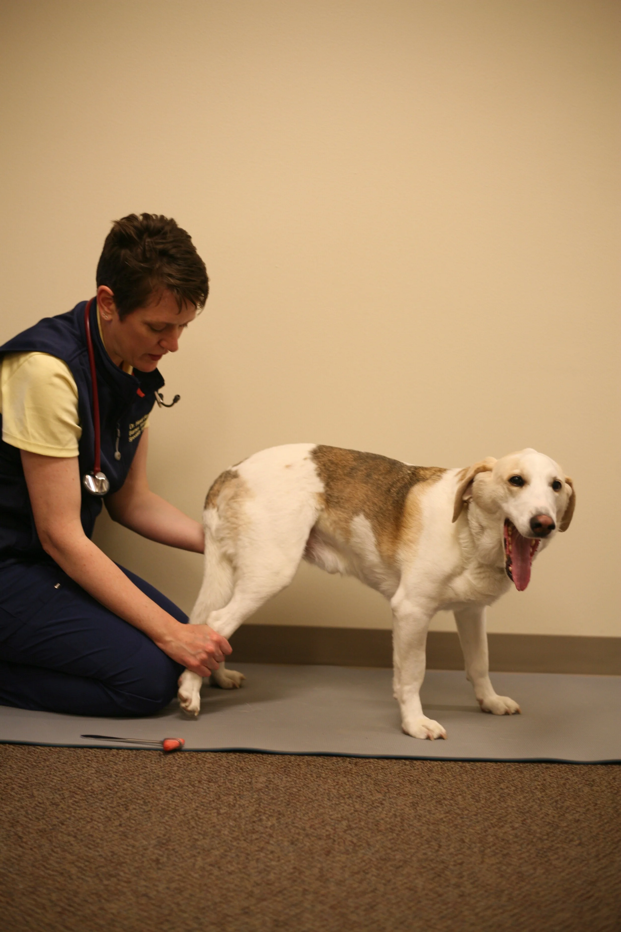
Performing CPs, or the paw replacement test, is so important for lesion localization, or to even identify if a patient has a neurologic problem. But it is difficult! This test causes residents to fret and frown because subtle differences in how you hold a patient, the flooring, and even environmental stimuli can affect how they place their limb. If you practice…it gets easier!

How do you identify increased intracranial pressures following head trauma?

How do you identify increased intracranial pressures following head trauma?

Are you ready to evaluate your patient for increased inttracranial pressure? Read on to see what simple techniques you can use to find this deadly result of traumatic brain injury. Spoiler alert - You need a stethoscope, a blood pressure measurement and a mini-neuro exam!