CBD Treatment for Resistant Epilepsy in Dogs

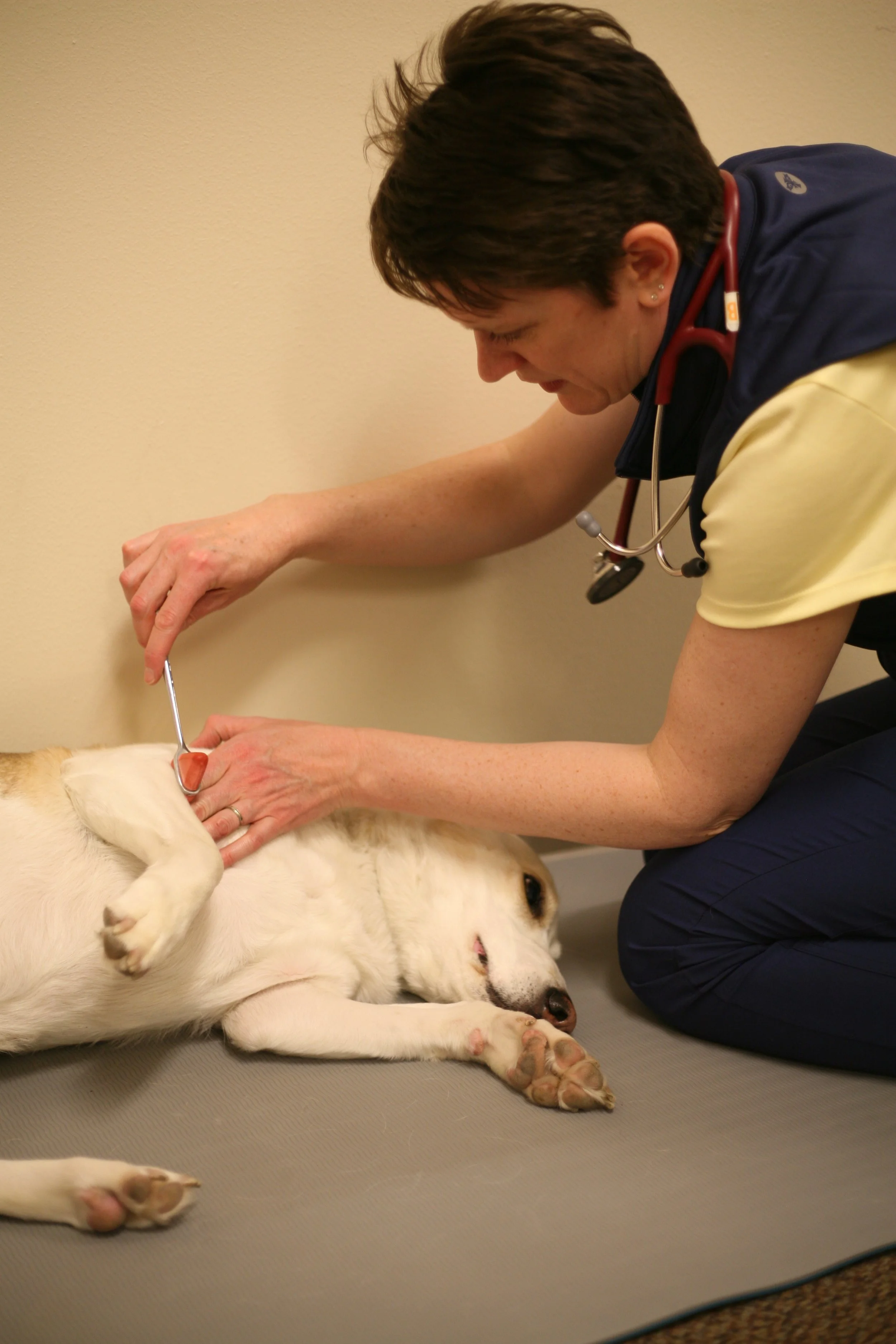
Use of CBD Oil for Refractory Epileptic Dogs


It's baaaack! At the ACVIM conference a few weeks ago I had the pleasure of listening to researchers from Colorado State University present data from a recent double blinded, placebo controlled prospective study evaluating CBD oil in resistant epileptic dogs. 

How the Study Worked

51 client owned dogs, taking at least 1 ACD (anticonvulsant drug) and continuing to have 2 or more seizures every month, were included. Resistant epileptics are already the hardest subset to manage so this was a really tough crowd for the researchers to target. The dogs were given either placebo or CBD oil for 3 months and then switched and given the other compound for 3 months. Initially, they dosed at 2.5 mg/kg PO q12h but no effect was noted so after 12 dogs the dose was increased to 4.5 mg/kg PO q12h. 

Key Points

  • ALP and ALT were elevated in this study. This is the first study to document elevated ALT during CBD administration in dogs. The researchers aren't sure why this happened but hepatocellular damage of some sort was suspected. Bile acids were normal in all dogs except 2.

  • No significant difference in response (response = a > 50% reduction in seizures) was noted between the placebo and CBD administrations group.

  • Total number of seizures/month and seizure days/month was reduced in the CBD group compared to the placebo group BUT over all seizures went UP with both groups

Should we use CBD to manage seizures for resistant epilepsy?


Sorry, not based on this study. It's not compelling enough to suggest that we should be using this drug PLUS it showed evidence of liver enzyme changes that could be concerning AND phenobarbital serum concentration went by 11% when the dogs were on CBD. That said, they targeted resistant epileptic dogs so could this be effective in "easier" to control dogs? Maybe. Could this increased phenobarbital concentration mean that we could lower the dose of phenobarbital in these pets? Maybe. Do we need to check out different doses? Maybe - but at least with this question I am cautious because of the new elevation of ALT at this higher dose. Can we safely increase the CBD dose? Unknown. 

This is a good step forward but alas, we still don't have enough compelling evidence to say CBD should be a part of seizure therapy and at what dose.

Keep up the good work managing epileptic dogs! I'm available if you have questions about a case or need a second opinion. Have a great week!

What is Temporal Lobe Epilepsy?

Temporal lobe epilepsy (TLE) is a newly emerging term in feline seizure discussions and one worth becoming acquainted with. TLE is not an etiology, but rather a specific seizure phenotype (appearance) caused by epileptic seizures in a specific part of the brain (hippocampus) resulting from multitudes of causes.

What does TLE look like?

Cats with TLE have 6 specific stages or appearances. Not all cats go through all 6 stages, nor are all 6 stages apparent. The stages are:

Stage 1: looking around, sniffing, attention

Stage 2: Immobility and staring (arrest)

Stage 3:Orofacial automatism (lip smacking, facial twitching, swallowing, blinking) hypersalivation, mydraisis

Stage 4: Masticatory movement, facial twitching

Stage 5: Head turning, head nodding

Stage 6: Generalized convulsive tonic-clonic seizure

Most cats are observed in stage 2 or 3, and may progress to stage 4 and 5 but stage 6 is rarely observed.


What causes TLE?

Because TLE is not an etiology, but a description of a seizure syndrome located in one part of the brain, any cause of non-TLE seizures can cause TLE seizures as well. This list could include vascular causes, neoplasia, meningoencephalitis (infectious or non-infectious), or idiopathic.

How is TLE diagnosed?

When orofacial automatisms are observed, TLE should be suspected. To confirm TLE, an MRI with changes in the hippocampus is needed. There may be evidence of the seizure etiology such as neoplasia, or vascular disease as well, but the hippocampus must show specific MRI changes. Histopathology can reveal hippocampal sclerosis or necrosis. Some authors think sclerosis precedes necrosis but this sequence isn’t fully established. I won’t bore you with the detailed differences between sclerosis and necrosis, especially since the difference is detected on necropsy.

Can we treat TLE?

Yes! Treat seizures with antiepileptic drugs (AED) as you would any feline or canine seizure disorder. Any 1 seizure more than every 3 months, or longer than 3 minutes warrants a discussion of starting AEDs. Recently, hippocampectomy (removal of part of the hippocampus) has bene published in a cat as a treatment for TLE. This is a well-established treatment for human TLE (a common form of AED-resistant epilepsy in humans) and it carries an extremely high success rate for seizure elimination. Unfortunately, the procedure in cats needs a bit more work before we start considering it standard treatment. The hippocampus is the memory storehouse therefore loss of the hippocampus can cause difficulty with motivation, spatial memory, and learned behavior. Some researchers have described the effect to be similar to feline cognitive disorder in geriatric cats. Hippocampectomy was extensively performed in the 1960s during the time of rapid, although sometimes perhaps unethical, knowledge acquisition of how the brain works in animals and humans. We must acknowledge this work, even if we might find it ethically repulsive today.


Key Points

·         Temporal lobe epilepsy is a syndrome, resulting in damage to the hippocampus and a classic seizure presentation.

·         Temporal lobe epilepsy can be treated, but may be progressive and unresponsive to treatment once the necrosis stage has been reached. How do we know we reached this stage? No one knows antemortem….stay tuned!

·         Cats should take phenobarbital or levetiracetam. These are currently (2023) the only two drugs with enough literature to support their use and predict a reasonable efficacy. (Sorry my zonisamide-using friends.)

Intervertebral Disc Herniation in Yorkshire Terriers


Yorkies are a popular, and common dog breed in the USA and I (we?) see them quite a bit in neurology referral practice. That said, I had never read that they were officially considered a chondrodystrophic breed. Apparently, they are! 

Intervertebral disc herniation (IVDH) occurs with high frequency in chondrodystrophic dogs, but how often do we see it in Yorkies? According to a recent article, about 10% of Yorkshire Terriers with neurologic disease are diagnosed with IVDH and undergo surgical intervention. 

How do Yorkies present with IVDH?

  • Cervical hyperesthesia only 5/60 (8.3%)

  • Ambulatory tetraparesis with or without neck pain (grade 2):  26/60 (43.3%)

  • Non-ambulatory tetraparesis or plegia with or without neck pain: 29/60 (48.4%)

No association with recovery and presence or absence of ambulation was found in this study. This is inline with other studies that have not found voluntary motor to be a prognostic indicator. 

  • Acute signs in 80% of dogs

  • Chronic signs in the remaining 20%

Yorkies with IVDH instead of another neurologic disease were statistically heavier and older

How do Yorkies do with surgery?

Thankfully, quite well, according to this report. In this study, the majority (82%) had one IVDH, 15% had two and 3% had 3 IVDH site repaired. In my experience, Yorkies far exceed dachshunds for having multiple IVDH requiring surgery at the time of diagnosis. Could this be a difference in genetic pool (this study was conduced in Czechia, Slovakia and Hong Kong)? In this study, all dogs returned to ambulation at some point, post operative. Most were walking by hospital discharge, but not all. This is comparable to dachshunds. 

Key Points

  • Yorkshire Terriers are chondrodystrophic and therefore predisposed to type I IVDH

  • Surgical intervention is likely to improve ambulation

  • Approximately 50% of Yorkies will be ambulatory, and 50% will not at the initial diagnosis. 


Thanks for reading! I am leaving for ACVIM today (Tuesday June 13th) and will be out for the remainder of the week. I'm so excited to collaborate with colleagues and bring new knowledge back to the patients we share! If you need me, email is preferred but texting is also fine. I will have limited ability to answer the phone but will do my best to return your call in a timely manner. Please excuse any unusual delays! Have a great week!
 

Immune-mediated Polyneuropathy in cats

Riddle me this: a 10 month old cat is brought into your exam room by a very concerned owner. They report that the cat has had a rapid, progressive weakness develop over the past 2 weeks. Being the astute veterinarian that you are, you notice that the cat takes 3-4 steps and then sits. There doesn't appear to be any ataxia...just paresis (weakness). 

Neurologic examination:
Mentation: BAR
Cranial nerves: all normal (about 11% of cats will have facial nerve paralysis with this disease)
Gait: Ambulatory tetraparesis, worse in the pelvic limbs
Reflexes: reduced withdrawal all limbs, absent patellar reflexes bilaterally. 
Postural reactions: reduced to absent tactile placing in both pelvic limbs, reduced tactile placing in both thoracic limbs. 
Palpation: non-painful

The remainder of the physical examination is unremarkable.

What is the neuroanatomic lesion localization?

Start at the very beginning. Are there any signs of seizures, mentation changes or cranial nerve deficits? No. Therefore this cannot be intracranial lesion localization. We have all four limbs affected therefore if this is a spinal neuroanatomic lesion localization it must be cranial to the front legs. This means C1-C5 or C6-T2 myelopathy. (If the lesion was T3-L3 or L4-S3, the thoracic limbs should be normal.) To narrow it down between C1-C5 or C6-T2, we look at reflexes. Reflexes are reduced in the thoracic limbs therefore this would suggest a C6-T2 myelopathy. BUT WAIT!! The reflexes in the pelvic limb are also reduced to absent and that cannot happen with a C6-T2 spinal cord lesion. The reflexes in the pelvic limbs should be normal with a C6-T2 spinal cord lesion. Is this a multifocal lesion, affecting two spinal cord segements (C6-T2 and L4-S3)? Although possible, you would expect a proprioceptive ataxia with a spinal cord lesion therefore this is very unlikely to be a spinal cord lesion at all, even a multifocal spinal cord lesion. There, we must assume this is not spinal cord or intracranial lesion. What does that leave us? Neuromuscular disease, that's right! Your options are: 1) peripheral nerve, 2) neuromuscular junction or 3) muscle. Animals with muscle disease should have normal reflexes (and do, except with myasthenia gravis), so it isn't that. This is either a neuromuscular or peripheral neuropathy localization

Our differential diagnosis would include diseases such as: toxoplasma or fungal neuropathy, myasthenia gravis (yes, I know this localizes to the muscle but clinical signs look like a junctionopathy at times!), and immune mediated neuropathy. 

Diagnostic testing for this patient

CBC, serum biochemistry (including CK!) is normal. Toxoplasma testing was negative, as is the myasthenia gravis titer. Occasionally we will see an elevated CSF WBC or protein count with this mystery disease, but it isn't common. 

Muscle and nerve biopsy results

Diffuse variability in muscle fiber size is expected on biopsy. Nerves are almost all missing from the muscle biopsy sections. The nerve biopsy is markedly abnormal, with inflammatory cells infiltrating the axon, nodes of Ranvier and Schwann cells. 

Diagnosis: Immune mediated polyneuropathy!

This is a very interesting disease for many reasons but most notably, most cats can achieve a complete recovery without any treatment at all. In a recent study (see below), 10/11 cats that received no treatment obtained full recovery with a median time of 4 weeks. Nine of 12 cats that received glucocorticoids and 18/20 cats that received L-carnitine supplementation recovered as well. Relapses are reported and can occur months or years after resolution of signs. Overall, all except 1 cat improved regardless of treatment in this study. This same study found a predominance in males but did not find a breed association. 

So there you go! A self resolving, relapsing remitting polyneuropathy of young cats. 

Article: DOI: 10.1111/jvim.16701

I hope you have a great week and look forward to working with you soon!

Long-term infection and monitoring of Brucella suis in dogs

B.suis, you say? Why do I need t know about a swine variant of Brucella?? Did you know that B. suis can be pathologic in dogs, as well as swine, and is also zoonotic? You're welcome :)

A recently published article detailed the clinical signs, blood work, imaging (when available), serologic progression over time, and body fluid levels of organism over an 18 month period in 27 dogs from 17 households. The study population was located in Australia where naturally occurring B. canis is not found. Exposure to feral pigs was considered an important source of disease acquisition and may be a stand alone reason to test. (1 in 10 pigs are positive in Australia.) Although B. suis is not as commonly noted in our US canine population, we are a global veterinary profession now with lots of dogs traveling or being adopted from international locations. I felt this article was a nice reminder that B. suis exists, and is pathologic in dogs as well as having zoonotic potential, like B. canis

Clinical Signs of B. suis

Ten dogs had clinical signs attributed to brucellosis. Clinical signs included pyrexia with orchitis and epididymitits (4/10), lymphadenopathy (1/10), spinal pain (3/10), abortion or dystocia ( 1/10), and athritis (2/10). The remaining 17 dogs were assymptomatic throughout the entire 18 month period!


Serology and PCR

Titers did not correlate with severity of signs. Postive results were deemed positive if a Rose Bengal rapid plate test (RBRPT) yielded a positive result along with a complement fixation test (CFT) greater than or equal to 8. Only 2 dogs had seronegative results after 18 months and both had low baseline serology. Brucella DNA was not detected by PCR in blood, urine, saliva or preputial swabs in any dog at any time in the study. Notably, they weren't swabbed weekly or daily, so it is possible that some shedding occurred without detection on the sporadic testing. Only 3 positive PCR tests were found and this occurred in 1 bitch post whelping (vaginal fluid), as well as milk at 3 and 7 weeks post whelping as well as a lymph node at necropsy on a puppy that died. Although culture may be the gold standard diagnostic test for some countries, the RBRPT and CFT were considered safter to personnel performing the test and were preferred in this study. 

Diagnostic Imaging Results

Fifteen dogs had some sort of radiograph taken during the study period. Two of 15 had joint siease, 3/15 had vertebral lesions and 2 of 15 had lymphadenopathy. The dogs with radiographic changes had spondylosis, which we know can be a normal finding, therefore I question the inclusion of this as a clinical sign of brucellosis. 

Treatment Response

Six dogs were treated with doxycycline and rifampicin (one dog relapsed and was treated twice. It was generally well tolerated with one case of marked hepatitis. There is no standardized treatment for brucella and many dogs are considered to harbor the bacteria long-term, with or without clinical signs, with intermittent bacteremia. The zoonotic risk to humans, therefore remains present with dogs with a current or historic positive Brucella test. Euthanasia is the recommended treatment for positive livestock, however this causes emotional conflict for veterinarians and owners of companion animals. Check with your local veterinary health directors to determine the legal requirements in your state or country. This is a reportable disease in the USA. 

If you'd like to read more about this study you can find it here: https://onlinelibrary.wiley.com/doi/epdf/10.1111/jvim.16678

In honor of Memorial Day yesterday, I wish to thank all of you that have served or have friends or family members that have been lost during military service. We honor you!


Have a wonderful week! My schedule has changed for summer so please reach out if you cannot find a suitable time for a consult online - I may have additional flexibility that isn't listed. 

Seizures, Quality of Life and Side Effects, oh my!


A study published in JAVMA (Gristina BR et al. JAVMA 2023)  just recently caught my eye and I thought, perhaps it might interest you as well. The study evaluated 100 dogs with , with Tier I or Tier II level confidence of idiopathic epilepsy and assessed owner satisfaction, seizure control and adverse effects (the fancy name for side effects) of various drugs. 
*Tier I = normal CBC, serum biochemistry, neurologic examination and bile acid test
* Tier II = all of tier I plus normal brain MRI and CSF analysis

How was the Seizure Control?

Improvement in seizure control was reported in 86% of dogs with phenobarbital, 76% in the levetiracetam and 65% in the zonisamide group. Treatment failure, due to inadequate seizure control, was 48% for phenobarbital, 32% for  levetiracetam and 35% for zonisamide. Importantly, they didn't corelate seizure control with serum drug concentrations because we don't have target serum concentrations for levetiracetam and zonisamide. HTerefore, some dogs could have been under medicated, and thus poorly controlled. Interestingly, 88% of dogs were still on their original antiepileptic drug (AED) at the time this study was performed. Mean daily doses for phenobarbital, levetiracetam and zonisamide were 4.9  mg/kg, 53.8 mg/kg and 12.4 mg/kg, respectively. 

What side effects made a splash?

Phenobarbital had the highest reported adverse effects at 77%, followed by levetiracetam at 59% and zonisamide at 39% of dogs. The most common adverse effects are listed below for each drug:

  • Phenobarbital: polyphagia, polydipsia, ataxia, sedation and polyuria

  • Zonisamide: sedation, ataxia, hyporexia

  • Levetiracetam: Sedation, ataxia, hyporexia, diarrhea, behavioral changes.

What was the perceived Quality if Life?

Owners perceived a significant improvement in quality of life, regardless of the anticonvulsant used, between pre and post treatment assessment. This is important for us to realize: clients can tolerate adverse effects if they perceive improved seizure control and quality of life! Although this retrospective, owner-perception study has limitations (all studies do), I felt it was worth repeating a bit of the information for you to add to your knowledge when addressing seizure management with your clients. 

Have a wonderful week! Remember, early bird registration ends May 31st for the July CE event so register soon if you are planning to do so! Details are available on my website. 


Reference: doi.org/10.2460/javma.22.10.0469

How to Question an Owner

Careful questioning of the owner is required to determine if the episodes described ARE seizures. Syncope, vestibular signs, neck pain, and movement disorders (think Scottie cramp) have episodic presentations with similarities to seizures. Nothing is fool proof, even an EEG, but here are some tips to get you going in the right direction. 

Describe the event, please!

Discrete episodes, with a finite start and stop, combined with autonomic signs often indicate a seizure. Level of mentation can be confusing and difficult to determine (especially for those pesky night time seizures) so don't spend too much time grilling an owner on this one. Videos can be priceless. I have evaluated many dogs and cats for seizures that actually have something else after reviewing the video. Neck pain, movement disorders, and syncope are the most common pretenders that I've seen. Finally, ask how long the events are lasting. This question is subject to tremendous bias, but if the owner says "all day" I start wondering about other non-seizure events. 


How often have the events occurred?


Okay sorry, I need to harp on this one. My pet peeve is hearing "about once a month" as an answer! This is an easy one and something we should encourage ALL clients with seizure pets to do. Keep a calendar! Tell owners to write it down, put it in a spreadsheet, mark it on their phone, keep a list - the choices are as varied as the seizures they record! You will NEED this to be in place to help you direct treatment. The single biggest reason to change treatment is that the seizures do not meet the seizure goals for epileptic pets. What are the seizure goals? Animals should have 1 seizure or less every 3 months. 

Your second goal here is to learn about cluster seizures. If the animal has 2 or more seizures in 24 hours that is defined as cluster seizures. Cluster seizures need at home cluster seizure management (another topic for another day). Furthermore, some drugs work better for dogs with cluster seizures than those with single seizures. I personally believe that bromide is a terrific option for cluster seizures and will readily use it for patients with this type of pattern. 

How long is each seizure? 

This question is utilized to learn about status epilepticus. Any seizure longer than 3-5 minutes (people argue about what is the correct time) is considered status. Status warrants emergency management with injectable solutions (intranasal, intravenous, other). Untreated status can set an animal up for systemic side effects as well as increase the risk of permanent brain damage. 

What does the animal do after the event is finished?


Your goal here is to evaluate the post ictal phase so that you can decide if a change in treatment is needed. Based on the rules outlined by the International Veterinary Epilepsy Task Force, severe post ictal changes (such as aggression) warrant treatment even if the other parameters for treatment haven't been met. I also use this question to determine how the owner is feeling about the event. Answers such as "he was fine" or "he paced and paced and seemed really upset" give me a window into how they feel about as much as how the dog did. Helping owners cope with seizures is also part of our job!

Do you have a seizure case that isn't meeting your seizure goals? Let me know! My favorite part of neurology is long-term seizure management so I'd love to help you, help your patients!

I'm headed to Chicago Vet Conference on Friday for a few talks on seizures, the neuro exam and lesion localization. I'd love to see you if you're at the conference so stop by and say "hi" if you have time!

Pituitary Apoplexy in Dogs

Pituitary Apoplexy in Dogs

What is Pituitary Apoplexy and What Does It Look Like?
Pituitary apoplexy is a clinical diagnosis caused by acute hemorrhage or infarction of the pituitary gland. The hemorrhage occurs secondary to neoplasia (benign or malignant). The hemorrhage is thought to occur because the tumor grew faster than the blood flow, or because of compression of the very sensitive blood vessels in the area of the pituitary gland.  Apoplexy isn’t common with pituitary neoplasia, and rarely fatal, but remains an important possibility for patients with pituitary gland neoplasia. Clinical signs described on presentation for dogs with pituitary apoplexy include acute onset mentation changes (obtunded, stupor, coma:62%), cranial nerve deficits (65%), gastrointestinal signs such as vomiting or nausea (54%), gait changes such as circling, weakness/paresis, ataxia (85%), and hyperthermia (31%). Other signs such as bradycardia, and cervical hyperpathia were noted but less commonly. The GI signs are critical, and unique, and shouldn’t be ignored. They could be due to vestibular signs (present in about 15% of dogs), but also could indicate a rapid rise in intracranial pressure.
Do all Dogs with Pituitary Apoplexy have Endocrinopathy?
No! According to a recent study by Woelfel et al, only 50% of dogs with pituitary apoplexy diagnosed on either post mortem exam or presumptively diagnosed on MRI had an endocrinopathy. A further 12% had signs suggestive of an endocrinopathy but did not undergo workup. This means that 38% of dogs did not have clinical or biochemical evidence of an endocrinopathy. The endocrinopathies could be the obvious one (Cushing’s disease), or the less obvious (central diabetes insipidus, hypothyroidism).
Treatment and Survival
Dogs receiving radiation therapy survived longer than those medically managed. There is probably a bit of bias, however, because the severity of clinical signs likely steered clients or veterinarians towards or away from treatment. The use of hyperosmolar solutions (mannitol, hypertonic saline) was associated with a poorer survival. Again, this may be due to the severity of signs of those patients receiving this treatment rather than the treatment itself. No clinically useful markers were identified to predict survival in this study but that doesn’t mean they don’t exist; just stay tuned!
Take home message: Acute onset mentation changes with vomiting? Get those dogs to a neurologist (or get a consult!!) as soon as you can.

I hope you have a wonderful week! I look forward to seeing you soon!


Reference: https://doi.org/10.1111/jvim.16703

Trace Minerals and Canine Epilepsy

Trace elements are the micronutrients found throughout live animals that are essential for organ function and brain health. They keep mitochondria running smoothly, improve neurotransmission and aid with enzyme function. Deficiency or excess has been linked to multiple neurologic diseases including neurodegenerative diseases, behavior diseases and inflammation in humans and animal species. A recent study evaluated the levels of several trace elements in hair samples of dogs with epilepsy and compared them to dogs without seizures.
 

What Trace Elements were Abnormal?

In this study, by Rosendahl et al, there were 10 epileptic dogs without any treatment, 53 epileptic dogs currently undergoing treatment for epilepsy, and 42 control dogs (no seizure history).

  • Phosphorus: lower in epileptic dogs

  • Copper: Higher in epileptic dogs

  • Zinc: higher in epileptic dogs

  • Copper/zinc ratio: higher in epileptic dogs. Specifically, higher in dogs treated with phenobarbital, in one previous study. Studies have shown that copper homeostasis is an important preventative for some neurodegenerative diseases. More work needs to be done in veterinary species to determine if this holds true for our patients, too.

  • Selenium: higher in epileptic dogs

  • Arsenic: higher in epileptic dogs but also much higher in dogs receiving potassium bromide!

Some of these findings were significant, some were significant only when comparing control dogs with either treated, or untreated dogs, and others were significant for both subgroups of epileptic dogs.

This study is likely to be a stepping stone for either this group, or others, studying trace minerals and their relationship to seizures. It was important work to identify differences, but the clinical significance remains unknown. Treatment, or correction of these deficits or elevations of trace minerals has not been evaluated in epileptic dogs. Stay tuned!
 

Thanks for reading! I hope you have a great week and enjoy what I hope is our final push out of winter, into spring, weather.

 
Reference: https://onlinelibrary.wiley.com/doi/full/10.1111/jvim.16698?campaign=wolearlyview

Neurologic signs of Hypertension in Cats

Hypertension is common in cats. It can be associated with a predisposing disease such as chronic kidney disease or hyperthyroidism, or idiopathic. Idiopathic hypertension is diagnosed when all predisposing causes have been eliminated (and hypertension is documented on 2 separate occasions)  and accounts for less than 1/2 of the cats diagnosed with hypertension (about 40%). 

Target organs for hypertension are the CNS, kidneys and cardiovascular system. Despite under recognition by clients, several studies have suggested that hypertensive encephalopathy might be present in 30-40% of cats. 

Neurologic manifestations of hypertension in cats

Seizures, mentation changes, vestibular signs (central), behavioral changes (disorientation), tremors, sudden collapse, cervical ventroflexion, paraparesis, cerebellar ataxia with hypermetria, cranial nerve deficits and cortical blindness (not retinal blindness). That's quite a list, isn't it?? It's amazing that these signs are the reason for presentation to a vet in only 10-20% of cats! 

A small study was recently published in the Journal of Feline Medicine and Surgery that aimed to identify the clinical occurrence of hypertensive encephalopathy in cats. In this study, 31 of 56 cats were diagnosed with neurologic signs associated with hypertension. Retinal lesion were identified in 28 of the 30 cats that under went fundoscopy. 

Cats with neurologic signs presented most often with proprioceptive ataxia, some with vestibular ataxia. Additional signs included hiding, disorientation, sleeping in unusual locations, increased and inappropriate vocalization, and increased appetite. Three of the 31 cats had seizures. Neuroanatomic lesion localization was predominantly prosencephalon, second most commonly vestibular signs and lastly spinal cord signs. Many cats had multifocal neurologic signs. 

Treatment and Outcome

Amlodipine was used in 22/31 cats. Telmisartan was used in 4/31 cats. The remaining cats received combination therapy. Follow-up was available for 25 of 31 cats. Fifteen cats had complete recovery after starting antihypertensive medications including one cat with severe seizures. Partial improvement was noted in 8/25 cats with residual ataxia or seizures manifesting most commonly. No initial response to treatment was noted in 2 cats with subsequent euthanasia within 1 week of treatment initiation. 

Key points:

  • Feline hypertension is common (they enrolled 56 cats in less than 2 years!)

  • Neurologic and behavioral signs occurred in more than 1/2 of the cats in this study but clients weren't aware of the significance!

  • Treatment with antihypertensive medication can result in improvement

  • Routine monitoring for hypertension is recommended in at risk cats, especially those with neurologic signs!

Reference: https://doi.org/10.1177/1098612X231153357

Thanks for reading! Have a terrific week; I look forward to working with you soon. Remember to sign up for the July CE if you are planning on attending because space is limited!

Unilateral Masticatory Muscle Atrophy


It's a Wednesday morning and you see on your schedule a 7 year old dog with unilateral temporalis and/or masseter muscle atrophy. What parts of the neurologic system could be involved?

1. Muscle: A problem with muscle function, termed a myopathy, can result in muscle atrophy. The most common cause of temporalis and masseter muscle atrophy in dogs is masticatory muscle myositis (MMM), which is caused from an immune mediated attack against the muscle fiber. This is a UNIQUE form of muscle inflammation because the proteins on these muscles are embryologically unique (2M fiber type) from all other muscles in the body. We care about that because it means that we can identify an antibody (AB) test that we can run that is highly specific AND sensitive! What else should we consider? Infectious myositis, secondary to neospora or toxoplasma infection would be my second choice differential for many dogs with this clinical presentation. In this scenario, the protozoa get into the muscle, set up a secondary inflammation (myositis) and muscle atrophy results. Sometimes you can see a mild positive on the aforementioned MMM AB titer test, but it is low, if positive at all. However, if you concurrently test for neospora and toxoplasma (serum titers) you can catch this "false" positive, and treat the correct disease.

Treatment for MMM: Immunosuppressive steroids for 30+ days, followed by a taper protocol.
Treatment for infectious myositis: clindamycin or sulfa antibiotics until negative or stable titers. This is often MONTHS of treatment!

2. Cranial nerve 5: A dysfunction of CN 5 can result in denervation atrophy of the temporalis muscles. On the exam, look closely for concurrent signs of a sensory neuropathy to the face and if present, a CN 5 dysfunction should be suspected.  This might include reduced blink reflex, reduced or absent corneal reflex, and reduced response to nasal stimulation compared to the unaffected side. The differential diagnoses list is much bigger but typically centers around a few common causes (neoplasia, neuritis, hypothyroidism, and trauma). To diagnose a CN 5 neuropathy the best approach is a thyroid panel,  CBC, serum biochemistry and then a brain MRI followed by a spinal tap, along with titers for infectious diseases (as indicated). Not all of those tests are needed for each patient so pick and choose as appropriate for your case. 

Treatment for CN 5 deficits: this varies and is dependent on the underlying cause. It is a bit hard to summarize TidBit Tuesday-style. :) 

These cases can be puzzling to sort out so please reach out if you feel a neurologic examination is helpful for your patient. Have a great week!

Cervical Disc Herniation Associated Myoclonus in Dogs

Intervertebral disc herniation (IVDH) is the most common cause of cervical pain in small breed dogs. The most common clinical presentation is cervical pain with a normal neurologic examination, however in a few dogs gait deficits, paw replacement deficits, or reflex deficits can seen. Myoclonus, or a sudden onset, repetitive muscle contraction is seen in about 4% of dogs in a recent study from France. This muscle contraction is frequently confused for seizure behavior by clients so be on the look out for it! The classic presentation is a small breed dog that stops an activity, demonstrates myoclonus, and then resumes it's activity. Other signs of cervical pain (yelping, low head carriage, reduced range of motion) are often present when clients are questioned, so be sure to ask! 

What is the Significance of Cervical Myoclonus with IVDH?

The presence of myoclonus did not change the prognosis or outcome for the 20 patients in the recent study (JAVMA 2023: 261:4: 511-516.). Surgical correction resulted in less recurrence of signs, and immediate resolution in the post operative period compared to medical management. Approximately 25% of of medically treated dogs experienced another episode of myoclonus considered to be distinct from the original presentation. Medical management consisted of NSAIDs, gabapentin and, for some, tramadol. 

What is the Take Away?

  • Myoclonus can occur with mechanical or chemical irritation of cervical nerve roots 

  • Myoclonus does not affect prognosis

  • Surgical management remains the recommended treatment for rapid resolution of signs of pain and reduction in relapse/recurrance

  • French Bulldogs were over represented in this study!! (Again - See TidBit Tuesday in March for the list of Frenchie spinal cord diseases


As always, thank you for reading! I am thrilled to see the lovely weather on the horizon this week and hope you have a chance to enjoy some of it, too. 

Canine Myasthenia Gravis Update

What is myasthenia gravis?

Myasthenia gravis (MG) is a neuromuscular disease caused by congenital or acquired causes. Acquired MG is caused by an attack against the acetylcholine receptor on the muscle or  the muscle specific kinase (MUSK) protein. Acquired MG will be the focus of this TidBit today. 


How does myasthenia gravis present clinically?

Focal, generalized and fulminant forms have been described. Focal MG results in weakness of 1 or more muscle groups (ex: esophagus or pharynx) and does not cause limb weakness. Generalized MG results in limb weakness, with or without (usually with!) megaesophagus and pharyngeal weakness and the fulminant form causes acute severe flaccid paresis or paralysis often requiring hospitalization and intravenous intervention. 


How is myasthenia gravis diagnosed?

Acquired MG is diagnosed by a combination of clinical signs, positive acetylcholine receptor antibody titer and/or positive response to edrophonium challenge. A positive acetylcholine receptor antibody titer is considered the gold standard diagnostic tool.  Seronegative MG is reported in about 2% of cases and likely represents dogs with immune attack against the MUSK protein rather than the acetylcholine receptor. Edrophonium is a short acting acetylcholinesterase inhibitor that can be used in the preliminary stages of diagnosis. Animals are administered a small dose of edrophonium and monitored for clinical signs of improved strength. This test is not advisable for use in obtaining a diagnosis of focal MG. It is important to note that dogs with other neuromuscular diseases may have a positive edrophonium challenge therefore it should not be used as a sole diagnostic tool. 


What is the treatment for myasthenia gravis in dogs and cats?

Acquired MG is most often treated with an oral, long acting acetylcholinesterase (pyridostigmine). Signs of overdose are similar to signs of acquired MG (SLUD plus weakness) therefore it is recommended to start at a lower dose and slowly increase the dose over 1-2 weeks until clinical signs resolve. Immunosuppression has been used for pets with focal or generalized MG however the risk of secondary pneumonia is higher with immunosuppression and the clinical improvement does not appear to be better than with pyridostigmine administration therefore it is rarely recommended. 
Acquired MG will often result in remission within 8 months of the initial diagnosis. If remission is not achieved, follow-up testing for concurrent diseases that could trigger MG is recommended. This includes a thyroid panel (for thyroiditis), chest radiographs and abdominal ultrasound for a cancer screening. 

Thanks for reading! I hope you have a great week and I look forward to working with you soon. 

Cushing's Induced Muscle Weakness

Hyperadrenocorticism (HAC) or Cushing’s disease is a common cause of muscle weakness. Biopsies have shown that type II muscle fiber atrophy is common and clinical signs have included weakness in all four limbs, along with muscle atrophy. A less common form of HAC myopathy is a severe muscle stiffness (SMS) that results in a stiff or stilted gait. A recent study evaluated the signalment, presentation and treatment options along with long-term outcome in a collection of 37 dogs diagnosed with HAC.

What is HAC induced Severe Muscle Stiffness?

As the name suggests, the clinical manifestation of the unusual form of HAC myopathy includes stiffness of pelvic or all four limbs. The etiology is unknown but electrodiagnostic evaluation suggests a myotonia. Myotonia is defined as a sustained contraction after percussion (hammer reflexes) or voluntary contraction. The muscle will occasionally ripple visibly, but usually a prolonged limb stiffness can be seen. Dogs with HAC induced SMS have a ridged, stiff-legged gait when moving and don’t demonstrate appropriate joint flexion. According to Golinelli et al (DOI: 10.11.11/jvim.16620) over 60% of dogs had only the pelvic limbs affected at the time of diagnosis, the remainder had all four limbs affected. An elevated CK was noted in 75% of the dogs, suggesting muscle membrane breakdown or injury.

It was interesting to note that the diagnosis of SMS was more common in small breed dogs, weighing less than 15 kg. The time of SMS diagnosis varied in reference to the HAC diagnosis.  Almost 62% were diagnosed 1 month to 3 years after being diagnosed with HAC, 8% were diagnosed with HAC and SMS at around the same time and 30% were diagnosed with SMS 1 month to 1 year BEFORE being diagnosed with HAC. There are many reasons for these findings including owner or veterinarian awareness of SMS, lack of ability to test for or diagnose HAC, or perhaps a pathologic change/trigger required to convert from HAC with weakness to SMS.

How is it treated?

In short, poorly. In the report cited above, no single treatment resulted in effective resolution of the clinical SMS. Therapeutic intervention included benzodiazepine drugs (19% of cases), physiotherapy (16% of cases), acupuncture (5% of cases), and one or two dogs received mexiletine, NSAIDs, dantrolene, botulin toxin, L-carnitine, methocarbamol, gabapentin, and cannabinoids. Mild improvement was reported with some treatments. Survival of dogs with HAC induced SMS was unchanged compared to survival of dogs with HAC only.


Key points:

Be aware that SMS can occur with HAC

The reason a dog will have SMS instead of muscle weakness is unknown

No specific treatment resolved signs in all dogs, however there are several reasonable treatment choices listed above to try.


Thanks for reading! Enjoy the first day of Spring!

Cluster Buster Protocol

What is a cluster seizure?
Cluster seizures are defined as 2 or more discrete seizures within a 24 hour period. Cluster seizures are different than status epilepticus (any one seizure lasting for an extended period of time, or 2 seizures in which poor recovery occurred between seizures). Cluster seizures pose a special concern for seizure patients because of they have been linked with poorer outcomes compared to dogs without a history of cluster seizures. 


The goal with cluster seizure management is to minimize the number and duration of seizures. A secondary goal is to reduce the need for hospital visit or stay thus reducing the financial burden to the client. 

When should I provide a cluster seizure plan to a client?

  • If a patient has a history of 2 or more episodes of cluster seizures

  • If the patient has a history of status epilepticus 

What drug choices, routes of administration and doses can I offer?

Levetiracetam

  1. Rectal administration – 40 mg/kg per rectum (PR) at the time of seizure, repeated once if needed within 24 hours. Results suggest an improvement in seizure management in the medium term using rectal levetiracetam AND standard anticonvulsant therapy according to one study

  2. Oral administration – start 20-30 mg/kg PO q8hr (NOT Extended release) levetiracetam after a pet experiences the first seizure and has recovered well enough to eat. Continue q8hr dosing for 2 days beyond the pet’s last seizure. The levetiracetam is then stopped, and the pet's long-term anticonvulsant therapy is continued. Clinical experience suggests this approach will reduce clinic visits, number of seizures during the cluster event. However, clinical side effects such as sedation or ataxia may be present during the dosing period. Note: This approach should NOT be used if levetiracetam is part of a patient’s standard oral therapy for long-term seizure control. Suddenly discontinuing an anticonvulsant medication can lead to breakthrough seizures or status epilepticus.

Benzodiazepine
Diazepam has been the main acute anticonvulsant treatment for veterinary patients.

  1. Rectal -  Be sure to target the rectum, not colon, to avoid hepatic metabolism. Start 1-2 mg/kg PR. Compounded suppository formulations of diazepam have not demonstrated reliable serum diazepam concentrations in dogs and therefore are not currently recommended.

  2. Intranasal – preferred by some due to the ease of administration, and avoidance of the first pass effect of metabolism. Start with 0.5 mg/kg intranasal. 

Midazolam

  1. Intranasal – this is my preferred route and drug for at home benzodiazepine administration. Additionally, it was reported to be favorable compared to intravenous midazolam in 44 dogs.Start with 0.2 mg/kg intranasal. 

  2. Intramuscular – this route is favored by some but is not my recommended route of administration for clients at home in emergency situations. In a clinical situation, intramuscular administration can be effective when intravenous access is limited. Starting dose of 0.2 mg/kg is recommended.

  3. Rectal – do not use midazolam rectally due to erratic and unpredictable plasma concentrations at standard doses.

 
Phenobarbital
Patients receiving daily phenobarbital administration can benefit from an increase, or pulse, of phenobarbital therapy during a cluster event.

  1. Oral increase – in clinical practice we regularly recommend increasing a dog from q12h dosing to q8h dosing and continue this plan for 2 days beyond the last seizure. An example of this would be if a dog had a seizure Saturday morning. Upon recovery from the seizure an additional oral dose of phenobarbital (at the same oral dose administered) is recommended followed by q8hr dosing Sunday and Monday. Tuesday the dog would return to the standard twice daily dosing interval. This approach provides a slight increase of serum phenobarbital concentrations which may protect against further cluster seizures. Clinical adverse effects such as increased sedation, ataxia, polyuria, polydipsia, and polyphagia may become evident during the increased dosing period.

Summary
Providing options at home for cluster seizure care can reduce the need for hospitalization, and clinic visits. Additionally, any of the above treatments can be provided in hospital during seizure observation should that be needed. All the treatment choices discussed above are targeted for dogs and are rarely used in cats.

I hope your Tuesday isn't a cluster of craziness! As you well know, I enjoy helping you, help your patients with seizures live their best lives so please reach out if you're working through a case and need a little extra help. Have a great week!

Pug Myelopathies

Did you know that Pugs are commonly diagnosed with T3-L3 myelopathies? There are so many to chose from I thought I'd take this TidBit Tuesday and discuss some of the more common ones. 


Intervertebral disc herniation

Dis herniation is a common etiology of a T3-L3 myelopathy in many dogs, and Pugs are no exception. Type I and Type II disc herniations have been diagnosed in Pug dogs in the thoracolumbar region. Clinical signs include acute (or chronic if type II) variably painful (but often WITH signs of discomfort in this breed), pelvic limb proprioceptive ataxia followed by paraparesis. Signs can progress to paraplegia with loss of deep pain. Surgical and medical correction have been shown to be useful for Pug dogs and depend on the severity of the clinical signs.


Subarachnoid diverticulum

Subarachnoid diverticulum (SAD) can be described as cystic accumulations of fluid in the subarachnoid space around the spinal cord. Pugs are cited as among the most common breeds to be diagnosed with SAD. Clinical signs are slow in onset and often include incontinence early in the clinical course. Dogs with SAD are not typically painful. Medical and surgical management have been tried however neither approach appears to be an overwhelming success. Medical management is typically my preferred approach. 


Meningomyelitis and Neoplasia

Any and all breeds are at risk for these two diseases and Pugs are not overrepresented in this group. That said, clinical signs would include acute or chronic onset of signs with paraparesis and proprioceptive ataxia in the pelvic limbs. Treatment is medical for meningoencephalitis and medical, surgical or with radiation therapy for neoplasia. 


Degenerative Myelopathy

Degenerative myelopathy (DM) is caused by a genetic predisposition to demyelination of the T3-L3 spinal cord segments. Pugs are over represented and either confirmed or strongly suspected to have a mutation of the SOD1 gene (I cannot remember if it has been confirmed, yet!), like  German Shepherd dogs. Clinical signs include a slowly progressive, non-painful, proprioceptive ataxia that progresses to paraparesis, then paraplegia. If not euthanized, pets can progress to tetraparesis and eventually respiratory failure. A diagnosis is made through findings of a normal MRI and CSF analysis along with appropriate clinical signs. Genetic testing can be useful but isn't a stand alone test. 


Constrictive myelopathy

This is the new kid on the block. First published several years ago, constrictive myelopathy is caused by a fibrous band of tissue that circles the spinal cord and causes a constriction to CSF flow and compression of spinal cord tracts. Constrictive myelopathy is now thought to form due to a lack of development (hypoplasia) of the articular processes of the TL vertebra. If you imagine the articular processes are the "hands" that hold the vertebra together, supported by muscles and ligaments, becomes easy to see how hypoplastic articular processes might result in vertebral instability. This instability would then result in fibrotic tissue forming to help "stabilize" the joint. Sadly, this fibrous tissue then causes spinal cord constriction. To make matters worse, SAD can also form secondary to chronic spinal cord trauma so these poor dogs can have constrictive myelopathy secondary to hypoplastic facets and SAD in their T3-L3 spinal cord segments. Yikes. A diagnosis is made on MRI. A recent study (https://onlinelibrary.wiley.com/doi/full/10.1111/jvim.16639?) evaluated the incidence of constrictive myelopathy alone, or in combination with other spinal cord diseases in a group of Pug dogs. They found only constrictive myelopathy in 3 dogs, constrictive myelopathy combined with IVDH in 17 dogs and IVDH only in 9 dogs, IVDH + SAD in 2 dogs and articular process dysplasia in ALL 32 dogs!

Summary:
Pug dogs are at risk for many causes of paraparesis and proprioceptive ataxia. A detail neurologic examination, history , and appropriate diagnostic imaging and spinal tap can help determine the underlying etiology and subsequently direct treatment most specifically. 

I hope this TidBit doesn't give you the impression that I am anti-Pug - I adore these little dogs. BUT,  I recognize their predisposition to specific spinal cord diseases when assessing them clinically. Remember, not all dogs with pelvic limb weakness have a disc herniation! 
Have a great week!

Hypophosphatemia in Seizures Vs. Syncope

We've all been there, right? An elderly small breed patient with a grade II or III heart murmur presents to you with a history of "passing out". Clients aren't more descriptive than that, and they didn't catch a video. Is this syncope or was this a seizure?

You have several things to ask and look for (see prior TidBit Tuesday on syncope) but recent research has given us one more tool in our belt to help differentiate between these two diseases. 

Human patients with generalized tonic-clonic seizures demonstrate a reduced phosphate level within several hours of the event and patients with syncope do not. A study from Israel published data in January that strongly suggested a link between hypophosphatemia and seizures in dogs, as well. According to this study, 87 dogs with seizures and 26 dogs with syncope were included. Phosphate was evaluated within 3 hours of the "event" in question and noted to be decreased in 28 (32%) of dogs with seizures. None of the dogs with syncope had a low phosphate level. Dogs were excluded from the study if their creatinine was above the normal range to eliminate those cases with hypophosphatemia from renal causes. Median phosphate levels were significantly different between the two groups!

Key point: With an optimum cutoff value of 0.97 mmol/L (about equal to 3.0 mg/dl), evidence of hypophosphatemia has a 100% specificity and 44% sensitivity to diagnose seizures. 

Importantly, this tool should not be used as a stand alone finding to differentiate between seizures and syncope! However, identification of hypophosphatemia, within 3 hours of the witnessed event, could suggest a generalized seizure was more likely than a syncopal event if all other historical, physical examination and biochemical findings are concurrently evaluated. Important note: This has not been verified in dogs with focal seizures or complex partial seizures OR cats! 


This week's TidBit Tuesday is short and sweet. I hope you have a wonderful start to March 2023! 


Reference: https://doi.org/10.1016/j.tvjl.2022.105914

Horner's Syndrome and Cervical Myelopathies

Do any of you feel comfortable localizing Horner’s syndrome in a dog? If you do…skip the first section and read the data from a recent study about cervical myelopathy and Horner’s syndrome.  If not, please carry on and join us for an interesting look at Horner’s syndrome with cervical myelopathies.

 
First, Anatomy

The sympathetic pathway to the eye is a 3-neuron system. Neuron 1 starts in the thalamus, travels through the brainstem and cervical spinal cord to T1-T3 thoracic spinal cord segments where it synapses. Neuron 2 starts here and travels cranially, through the ansa subclavia along the vagosympathetic trunk (right next to that jugular vein you’re about to do venipuncture on!) to the caudal aspect of the bulla. From there, the 3rd order neuron takes a path through the tympanic bulla, along the ventral aspect of the skull (in the cavernous sinus) and hops a ride with CN 5 (trigeminal) to make a beeline to the eye. This neuron innervates the muscles of the iris, eyelids and orbit. It is the most indirect path anyone could design but I might argue that you can break it down into several key parts when localizing Horner’s Syndrome.

  • Intracranial

  • Cervical

  • Brachial plexus

  • Jugular groove

  • Tympanic bulla

  • CN 5

Cervical Myelopathies and Horner’s Syndrome

After reviewing the anatomy, it might be easy to see how a cervical lesion may cause Horner’s syndrome, right? The 1st order neuro travels from the intracranial structures via the cervical spinal cord to the upper thoracic spinal cord segments. Interestingly, a recent study looked at Horner’s Syndrome and cervical myelopathies* and found an incidence of only about 10% of Horner’s syndrome with concurrent cervical myelopathy. Therefore, although the anatomy makes sense, it is a fairly protected neuronal pathway and therefore a cervical lesion rarely causes Horner’s signs.

What Causes Horner’s Syndrome and a Cervical Myelopathy?

I’m glad you asked! According to this study, more dogs with Horner’s syndrome had noncompressive lesions compared to the control group (dogs with cervical myelopathy without Horner’s syndrome). Noncompressive lesions are often caused by fibrocartilaginous embolism (FCE) but hydrated nucleus pulposus extrusion (HNPE) and intramedullary neoplasia, noncompressive spinal trauma, Syringohydromyelia and inflammatory myelitis were found to cause Horner’s syndrome.  Also of note, Horner’s syndrome can be unilateral OR bilateral with cervical myelopathies.

Did having Horner’s Syndrome Affect Prognosis?

Nope. The underlying disease predicted prognosis and having Horner's Syndrome did not significantly negatively or positively influence outcome. However, seeing Horner’s syndrome on the neurologic examination would suggest that a surgical lesion is less likely to be identified.

 
Do you have a patient with Horner’s Syndrome? Do you suspect a cervical myelopathy? I’d love to help! Please reach out via email or schedule a consult online. Stay safe on these slippery roads and have a great week!

Reference from the TidBit Tuesday: https://onlinelibrary.wiley.com/doi/epdf/10.1111/jvim.16588

Bile Acid Testing For Dogs with Seizures

Case scenario: You are presented with a 2 year old Labrador retriever with a history of 3 seizures in the past 1 month. The seizures are consistent with generalized seizures and last less than 1 minute. Further questioning of the client reveals the dog to have normal activity, appetite, and mobility at home between seizures. You perform a neurologic examination (yay!) and no abnormalities are found. 


What is the likelihood of idiopathic epilepsy in this dog?

According to the International Veterinary Epilepsy Task Force, a diagnosis of idiopathic epilepsy can be made, at a Tier I level of confidence, if a dog is between the ages of 6 months and 6 years, has had 2 or more seizures, has a normal interictal neurologic examination AND has normal CBC, serum biochemistry and dynamic bile acid testing (that means pre and post feeding testing). We know Labs are commonly diagnosed with idiopathic epilepsy and that a genetic inheritance is known or suspected for most of the breed. So, do we really need to do a bile acid test? 

First, a little background. Minimum data base (MDB) pseudohepatic function tests include glucose, BUN, albumin, ALP and ALT. A pre-prandial bile acid test alone, called a resting bile acid test, is different than a dynamic bile acid test which includes both pre and postprandial samples. 

Do we Reallllly Need to Perform Dynamic Bile Acid Testing?

An article from England recently addressed this question in a publication in the Veterinary Record (DOI: 10.1002/vetr.2585).

Questions asked:
1. If  a dog has a normal MDB, how likely are we to finding an elevated postprandial bile acid test?" Answer: 24 dogs out of 202 dogs

2. How likely is a dog with a normal MDB and a normal pre-prandial bile acid test to have an elevated postprandial bile acid test? Answer: about 9 out of 100 dogs

3. What is the prevalence of a clinically significant hepatopathy in a dog with a normal MDB and normal pre-prandial serum bile acid test (if we don't do a post-prandial bile acid test)? Answer: 1.29%

The authors compared this to the risk of missing a significant brain lesion in a dog less than 6 years of age with a normal neurologic exam in which an MRI is not performed. (About 2.2% of cases would have had a brain lesion missed.) The question always begs, how much of a risk taker are you, or your client?

Based on the information from this study, here is what I propose we do:

  • ALWAYS check CBC, serum biochemistry for every dog with a history of 2 or more seizures.

  • ALWAYS recommend a pre AND post bile acid test for every dog presenting with a history of 2 or more seizures, even if CBC and serum biochemistry are normal. When making this recommendation I suggest that we make clients aware of the less than 2% chance that their dog could have a significant hepatopathy that will be undiagnosed if we do not perform these tests. This hepatopathy may be the reason for their seizures or, and perhaps more importantly, it could affect how they metabolize many of the anticonvulsants that we use. I'm looking at you phenobarbital, zonisamide and diazepam! Poor hepatic function could result in poor metabolism of these anticonvulsant drugs even if the hepatopathy isn't severe enough to be the seizure etiology. 

  • ALWAYS perform a neurologic examination to document any abnormalities before starting any medications for seizures. (Okay, so this wasn't part of the study but I still think this is a must!)

Thanks for reading! This was a very informative article so check it out for more detail! 

Have a seizure patient that you need a little backup for? Seizure management is my passion so I'd love to help! Email me or hop on my website to schedule a consult. Have a great week!