Anisocoria

** Credit for the amazing hand-drawn image goes to my good friend, and veterinarian Dr. Pam Boutilier.


Anisocoria

Anisocoria is defined as pupil asymmetry and may be seen with ocular or neurologic dysfunction. Malfunction of the sympathetic, parasympathetic, or visual system may result in anisocoria.

Neuroanatomy

Visual Pathway
When light enters the eye, it activates the light receptors in the retina. That information then travels along CN II, crosses in the optic chiasm, and terminates in the thalamus. Optic radiations relay the visual information from the thalamus to the visual cortex in the brain. This pathway can be tested using the menace response test and/or cotton ball testing.

Parasympathetic function: Pupil constriction
The parasympathetic pathway to the eye is a short, two neuron pathway which originates in the midbrain. The two, paired parasympathetic nuclei of CN III (PSNCNIII) send fibers along with the somatic nerves from CN III to the eye. The parasympathetic pathway is best assessed using PLR. When a bright light enters the eye, CN II activates and synapses on the PSNCNIII. The parasympathetic fibers transmit this information to the eye, using cranial nerve III as a conduit, resulting in pupillary constriction.

Sympathetic function: Pupil dilation
The opposing system is the sympathetic system, which causes pupillary dilation. The sympathetic pathway is a three neuron pathway and takes a longer course to the eye compared to the parasympathetic system. In general it goes from the thalamus, through the brainstem and cervical spinal cord to exit in the upper thoracic segments. Fibers then make a U turn and head back to the eye via the jugular groove (don't poke around too much when doing those jugular blood draws!), bulla (ear infection can = sympathetic dysfunction) and then hitches a ride with CN V to make the final leg to the eye. Malfunction anywhere along this pathway will result in a failure of iris dilation in a dark room and a comparatively miotic pupil, typically accompanied with enopthalmus and ptosis.

Putting it together

Let's put this new knowledge to work. If you see a case with anisocoria, how do you decide if it is a parasympathetic or sympathetic dysfunction?
1) Assess the pet in a dark room. Does the eye dilate? If yes --> The lesion is NOT due to sympathetic dysfunction.
2) Asses PLR. Does the eye constrict? If yes --> the lesion is NOT due to parasympathetic dysfunction.

To localize to the appropriate location beyond sympathetic or parasympathetic function requires a full neurologic examination. If you aren't comfortable performing or interpreting a neurologic examination please consider a neurology consultation! I am not comfortable doing a dental...we all have our limitations! :)
If you're interested in digging into anisocoria more deeply, or you have a case with anisocoria consider checking out the following article for a full discussion and more amazing images. Note: I do not receive any royalties or financial impact from this article.
* Barnes Heller H, Bentley E. The practitioner's guide to neurologic causes of canine anisocoria. Today's Veterinary Practice Jan/Feb 2016 pg. 77- 83.

Keep those consults coming; we all get to learn a little bit more with each consult. Have a great week!

Neospora caninum: Update!

Medical Review: Neospora Myositis

You might be wondering why on Earth I'm talking about Neospora as a small animal vet. (Neospora causes abortion in cattle!) It infects dogs too!! Some of you have seen some positive cases with me throughout 2020 so, I thought it was worthwhile doing a mini-review so we could all brush up on it together.

Etiology: Toxoplasma gondii and Neospora caninum may cause myositis, peripheral neuritis or CNS meningoencephalitis.

Signalment:

Breed: any
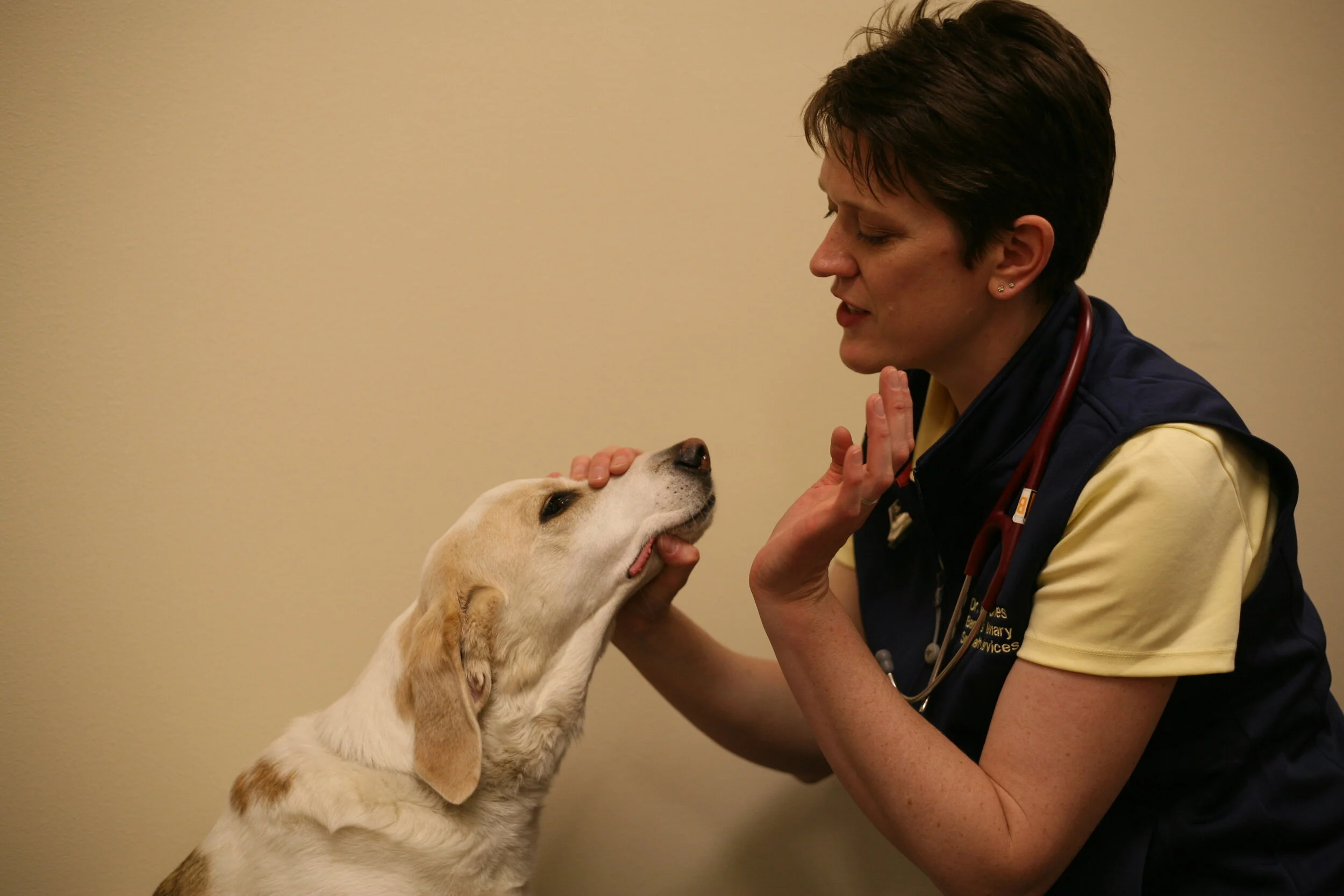
Age: may be acquired in utero leading to signs at a very young age (months), or by consuming raw or under cooked meat causing signs at any age. **Adult onset is what I see most commonly. However, the dog pictured above was a puppy that presumably acquired it in utero.

Clinical signs: Stiff gait, muscle pain (myalgia), muscle atrophy. Note: Muscle atrophy can be anywhere including the muscles of mastication. I have been tricked into thinking a dog has MMM when in actuality it had Neospora myositis causing unilateral muscle atrophy in the masticatory muscles.

Diagnostic tests: IFA > 1:200 for Neospora caninum is consistent with an infection, however documentation with a repeated titer is recommended as a confirmatory test. Muscle biopsy showing the encysted organism is definitive but obviously the most invasive route.

Treatment options: Sulfadimethoxazine/ormetoprim (15 mg/kg PO q12h) or clindamycin (10 mg/kg PO q12h) twice daily is recommended for 4-6 weeks, possibly longer. Be aware that sulfa antibiotics can cause idiosyncratic myopathic drug reactions too!

Prognosis: Good, if treatment is started before severe muscle atrophy or fibrosis is present. Prognosis is poor if clinical signs are severe, or chronic.

Frequency: Uncommon, but not unheard of!

Voila! TidBit Tuesday-style update on Neospora caninum. 😊

Final question: When do I run the IFA? Answer: Any dog with a history of muscle pain, muscle atrophy (not explained by neuropathic disease) or an elevated CPK should have a Neospora titer performed. This disease is sneaky so I err on the side of caution and suggest running the test rather than getting caught unawares.


Be safe, be strong and keep those referrals coming!
Heads up for those of you in my referral area: I will be closed Thursday January 28th to celebrate my youngest kiddo turning 5 years old. My, how time flies! I apologize for any inconvenience this may cause and look forward to consulting with you on another day!

Prognostic Indicators for Acute Myelopathy

Acute Paraplegia: When to ship for surgery, treat medically or consider euthanasia

It happens to all of us (perhaps to me, more than you). Monday morning, 9 am, we are presented with a dachshund (Shih Tzu, Corgi...pick your top chondrodystrophic breed) after the dog became acutely non-ambulatory over night.

You perform a neurologic examination (self high-five!) and determine that the dog has a T3-L3 myelopathy with spinal pain at the TL junction.

What do you do next?

To be clear, a T3-L3 myelopathy does NOT equal a type I disc herniation. It means the dog may have a type I disc herniation or it could have a fibrocartilaginous embolic myelopathy (FCEM), acute non-compressive nucleus pulposus extrusion (ANNPE) or in rare cases meningomyelitis. (Yes, even dachshunds can get meningomyelitis!)
If we play the odds, that chondrodystrophic dog likely has a disc herniation. What, if any, prognostic indicators can you provide to the client?

Thankfully, an article out of Frontiers in Veterinary Science entitled "Prognostic Factors in Canine Acute Intervertebral Disc Disease" was published in November 2020 for just this purpose! (1) Here are some key points from the article. (The entire article is available as an open access article if you wish to read it in it's entirety. I highly recommend doing so if you are in the habit of seeing paraplegic dogs.)

Prognostic Indicator Key Points:

  • Recovery following medical management (only) for dogs with paraplegia (loss of all voluntary motor) and loss of deep pain perception (DPP) is about 22.4%.

  • Recovery following medical management (only) for dogs with paraplegia and intact DPP is 56%.

  • Recovery following surgical management for dogs with paraplegia and loss of DPP is about 61%.

    • 25% recovered at 2 weeks

    • 42% recovered by 4-6 weeks

    • 53% recovered by 12 weeks

  • Recovery following surgical management for dogs with paraplegia and intact DPP is about 93%.

*** Did you catch that?? DPP is a prognostic indicator! Dogs without DPP do worse long-term compared to dogs with intact DPP. They are also have a 10x higher likelihood of developing progressive myelomalacia which is a fatal secondary outcome from acute spinal cord injury.


What exactly does "recovery" mean? It means walking without support but it DOESN'T mean walking normally or complete continence. Dogs may (and often do) have fecal or urinary incontinence consistently or occasionally following severe spinal cord injury.

Other Key Points:

  • Spinal shock can be more commonly associated with long-term fecal incontinence but does not seem to affect the ability to recover ambulation.

  • Age, breed and weight are not associated with prognosis...mostly. Some studies have found heavier dogs have a worse prognosis, other's haven't. My take - bigger dogs are harder to care for but that doesn't mean they cannot recover.

  • Reduced pelvic limb reflexes due to a L4-S3 lesion (not associated with spinal shock) has a higher likelihood of incontinence long-term.

When do you send a dog for surgery?

1) if the dog is non-ambulatory or paraplegic and the clients have a desire and financial ability to pursue surgery. Surgery with imaging costs $2000-5000 USD, depending on the specialty referral center.
2) Rapidly progressive signs (E.g.: dog was walking at 8 am, and is paraplegic at 10 am, with loss of deep pain by noon) and clients wish to pursue surgical intervention if indicated after imaging.

My plea...

Please don't send clients several hours to a specialist for surgery only to discover that the cost is prohibitively expensive. It is heart breaking for everyone and unnecessarily stressful for the dog. Call me and I'll happily talk with you about consult/medical management choices or sadly advocate humane euthanasia for the dog, if it is the best option.


Stay Safe, Keep Keeping On and Have A Good Week!


(1) Olby Natasha J., da Costa Ronaldo C., Levine Jon M., Stein Veronika M. Prognostic Factors in Canine Acute Intervertebral Disc Disease; Frontiers in Veterinary Science. 2020:7 p 913.

Monoparesis following Vehicular Trauma

How do you sort out a cat or dog with thoracic limb monoparesis following trauma?

This is an important question. First, let's review the innervation to the front leg. (Get back here - it's not that bad!) There are two important nerves that guide weight bearing and movement in the front leg:
1) Musculocutaneous - exits spinal cord segments C6-8. Important innervation is biceps muscle but it does a few others. The important action is flexion of the elbow and extension of the shoulder
2) Radial - exits spinal cord segments C7-T1 +/- 2. Innervates triceps and the muscles on the cranial distal limb that extend the carpus. The important action is extension of the elbow and carpus for weight-bearing.

So, to weight bear there must be an intact radial nerve. To move the limb forward there must be an intact musculocutaneous nerve. The other nerves (supra and subscapular, axillary, median and ulnar) are important too, but not as important. If you want to remember only two nerves, remember radial and musculocutaneous.

Now that we have that over with, let's put this to practice. Here is the scenario: You are presented with a 1 year old cat with a history of vehicular trauma a few days ago. The cat is presented dragging the left thoracic limb, unable to bear weight. When you watch it walk you can see advancement of the shoulder and elbow but it is minimal.
Question: What nerve is affected MOST?
Answer: Radial
Question: What spinal cord segment does the radial nerve arise from?
Answer: C8-T1 or 2.

Really good question: What is this cat's lesion localization?
Answer: Let's do a neurologic examination! :)

Neurologic exam:
Mentation: BAR
Cranial nerves: normal (note Horner's can be seen with thoracic limb injuries but isn't seen in this cat).
Reflexes: Absent triceps, absent withdrawal of the left thoracic limb. Unable to detect bicep or extensor carpi reflex (hey, it's a cat, give me a break!). All other limbs have normal withdrawal and pelvic limbs have normal patellar reflexes.
Palpation: non painful
Proprioceptive testing: absent tactile placing left thoracic limb, absent tactile placing left PELVIC limb, normal entire right side.
Gait: left thoracic limb monoparesis as previously described.

Now you can properly answer the previous question.
With absent radial nerve and diminished musculocutaneous innervation to the left leg, we KNOW the lesion must involve C6-T2, correct? (It's absent because of the missing withdrawal reflex - mediated through largely the radial nerve but also musculocutaneous too.) The real question here is this: is the injury in the plexus only or is there evidence of spinal cord involvement? To answer that question, you must look downstream from the affected segment. This means, look at the left pelvic limb. This limb has absent tactile placement so there has been disruption to the spinal cord tract going from the brain to the left pelvic limb, and back again.
Answer: This cat has a C8-T2 spinal cord lesion.

The differential list must now include things that affect the spinal cord such as avulsion and a disc herniation, hematoma or hemorrhage. Your diagnostic plan would include advanced imaging of the spinal cord. If the lesion localization had been peripheral plexus (not spinal cord) you would consider a brachial plexus avulsion only and advanced imaging would not be indicated. Knowing the lesion localization can markedly change your differential diagnoses, diagnostic plan and ultimately treatment and recovery!

Not sure about this case? Feel free to email me! This TidBit Tuesday is a slightly altered version of a real case seen recently. Keep those consults coming so we can share the knowledge folks! Please email/call/text me to schedule a consult or hop on my website and utilize the online scheduler to find the best time.


Have a good week!

Midazolam as a Constant Rate Infusion (CRI)


We Tell Clients: Seizures Should Be Stopped ASAP. But...Why?

  1. Prevent brain damage

  2. Prevent secondary systemic problems such as hypoperfusion, acidosis and possibly hypoglycemia if prolonged.

Okay, fine that's reasonable. But how, and when do I use a CRI?

My rule of thumb for dogs and cats with seizures is to recommend hospitalization with placement of an IV catheter and initiation of a constant rate infusion if a pet has more than 3 seizures in 24 hours. (At home cluster buster care is another TidBit Tuesday.) Once seizure control has failed at home, it is time for hospitalization.
Diazepam is the mainstay drug intervention however diazepam shortages and a growing recognition of phlebitis secondary to IV diazepam has lead to a greater use of midazolam. Full disclosure, I have used midazolam without hesitation for more than 12 years but...sigh...we didn't have scientific literature to support it's use, until now!
A study out of North Carolina State University by Dr. Bray and colleagues (two of whom were my resident mates way back...) described the use and safety of midazolam CRIs in dogs with status epilepticus and cluster seizures.

The findings, in a nutshell:

  • 106 dogs including 40 with idiopathic epilepsy, 16 unknown seizure etiology, 43 structural epilepsy and 7 with reactive seizures. --> I suggest that this heterogenous population provides us challenges with efficacy studies but at least we have something!

  • Median start dose was 0.25 mg/kg/hr., range 0.1-0.75 mg/kg/hr. (Remember it has a 20% higher binding affinity than diazepam)

  • Seizure control was obtained in 82 dogs (77% of the population) including 49 in which no dose adjustment was needed and 25 in which dose escalation was required to control signs and a further 8 in which 1 single additional seizure occurred but they still considered it control. (Hmmm, this group doesn't meet their criteria very well.)

  • Adverse effects were reported in only 24 dogs (22.6%) which included sedation, vomiting/diarrhea, hyperexcitability, ataxia and polyphagia. No reports of phlebitis, as expected.

  • Median time to control was 13 hours and CRI duration was 25 hours.

The main limitation to the study, from my point of view, is the heterogenous population. We suspect poorer seizure control in dogs with structural epilepsy than those with idiopathic epilepsy. This was also noted in the study wherein control was obtained in 85% of dogs with idiopathic epilepsy and 74% of dogs with structural epilepsy. This difference, however was not statistically significant.

Take Home Message

  • Continue to use midazolam, with a starting dose of 0.2 mg/kg/hr. (Now with data to support it's use!!)

  • Expect success in most patients, regardless of the underlying cause

  • Plan to use it for at least 12 hours, but possibly longer

  • Phlebitis is not expected, unlike with diazepam!

Want more? The full article can be found online (open access):
Bray, KY, Mariani, CL, Early, PJ, Muñana, KR, Olby, NJ. Continuous rate infusion of midazolam as emergent treatment for seizures in dogs. J Vet Intern Med. 2020; 1– 9. https://doi.org/10.1111/jvim.15993

Welcome to 2021!! I look forward to continuing to work with you, and your staff.

Seizure management is my passion - please call/email or have me out for a consult if you have a seizure case!

Happy New Year!

Happy New Year!
Good bye 2020 and hello 2021!

This year I have learned:

  1. I'm more resilient then I thought.

  2. My kids are fragile, and yet stronger than I gave them credit for.

  3. I love working, especially as your "pocket neurologist"!

Things I am looking forward to in 2021:

  1. Going swimming in a swimming pool (not a lake). I'm tired of seaweed...

  2. Playing with my children at parks without fear of their (and my) exposure to a potentially deadly disease.

  3. Traveling to a city in Wisconsin or Illinois that I haven't yet been to (come on Northern Woods vets!!).

  4. Hugging!!

  5. Continuing to work with all of you wonderful, dedicate, kind, strong and resilient veterinarians and veterinary staff. YOU are whom I look forward to when I go to work!

Wishing all of us a happy, safe and prosperous New Year!

The Nuts and Bolts of Anticonvulsant Drug Monitoring

The following information is contained in these two tables:
1. What drugs can I run therapeutic drug monitoring levels?
2. Where do I submit samples?
3. When do I draw blood for sampling?
4. What is the standard reference interval?
5. What is MY (i.e. Dr. Heidi Barnes Heller's) recommended reference range
6. How should I collect this sample? Note - all samples for therapeutic drug monitoring should be spun and separated into a plain red top tube. Do not use serum separator tubes! Plasma can be used for some samples also. Please separate the plasma into a plain red top tube as well.
7. What time of the day should I collect the sample?
*** The format became a little messy with the mailer so email me directly if you want a printable PDF of these tables.

What is not contained in these tables?
How do I use this information obtained from a drug serum concentration?? Stay tuned for that in a separate TidBit Tuesday! :)

As always, please stay safe, and mentally and physically healthy out there. I love working with you and look forward to continuing to do so.

Please note that I am performing consults through December 25th and then I will be modifying my schedule through January 16th because of my concern for increasing COVID numbers following the holidays. As many of you know, I have an at risk kiddo so I am super-duper cautious. If you need a consult between December 25th and January 16th please email me to discuss options.

Happy Hanukkah to those of you also celebrating!

20% of dogs receiving AEDs have dermatologic reactions??

Up to 20% of dogs taking AEDs may have a drug reaction that manifests as a dermatologic reaction.

Wow! That number is higher than I remember from residency; what's your experience?? I dug into this statistic this week after consulting on a case about a possible dermatologic drug reaction in a dog receiving an anti-epileptic drug (AED).

What signs are seen?

During a prospective study, 15 dogs (of 137 dogs) were identified as having a dermatologic condition after starting an AED. However, three may have had signs before starting the AED. All 15 were treated with phenobarbital!


9 dogs were seen by a dermatologist and the following signs were documented:

  • Extensive erosions or epidermal necrosis leading to skin detachment (4 dogs)

  • Papules, pustules, erythema, crusty lesions (8 dogs)

  • Pruritus and/or alopecia (3 dogs)

How do they *know* it is related to the drug?

They utilized an index based assessment, which is used for drugs in which withdrawal or drug challenges may be unethical (AEDs!). The index is called the Naranjo intext. I'm not terribly familiar with the nuances of using this index, so forgive my lack of a more detailed explanation. When they applied this, they considered 6 dogs to have probable cutaneous drug reactions, possible drug reactions in 8 dogs and doubtful in 1 dog. We all know these indexes can be wrong, but those numbers sound pretty compelling, and concerning.

How was it managed?

The AED therapy was withdrawn in 7 cases and 5 subsequently resolved completely with 2 weeks. Another dog had partial improvement after withdrawal of the drug and the rest received supportive or directed treatment such as anti-inflammatory, antimicrobial topicals, ant parasitic agents, or systemic prednisolone. All dogs had resolution of signs with the above treatments, but improvement wasn't complete and appeared temporary.

Key Point: Do a thorough physical examination on your patients before starting any AED and document new dermatologic changes if they arise. Can they still be coincidental? Yes. But, maybe we're are missing some that are not.

Do you need help with an epileptic patient? I am especially fond of helping with seizure management so I'd love to help! Call, text or email. Please note that I respond to emails at night (most days) so if you have an urgent question please call or text.

*Reference: Koch T, Mueller BD, et al. Cutaneous adverse drug reactions in dogs treated with antiepileptic drugs. Frontiers in Veterinary Science 2016: 3: 1-10.

Meningitis...Encephalitis...?

Canine Meningitis...Encephalitis...Meningoencephalitis... oh bother...

Canine Meningitis...Encephalitis...Meningoencephalitis... oh bother...

Canine meningoencephalitis is a general term, used to collectively describe inflammation in the brain and meninges of dogs. Although infectious meningoencephalitis can occur, the majority of dogs have a non-infectious, inflammatory disease.

Remember the term GME? Well, that is only ONE type of non-infectious inflammatory meningoencephalitis! Note: to call it GME we need histopathology. Not surprisingly, many clients do not opt for brain biopsies so we cannot confirm if it is GME, or NME or NLE or another histologic subtype of inflammatory brain disease antemortem. That won't do!

IF I CANNOT CALL IT GME IN A LIVE PATIENT, WHAT DO I CALL IT?


We call the collection of non-infectious, inflammatory brain diseases Meningoencephalitis of Unknown Etiology (MUE). Or, if you're British it's MUA (aetiology). Or, sometimes its called MUO (origin). Shoot, we're back to alphabet soup again. 😒 When we settle on a term, I'll let you know, but for now the literature typically refers to non-infectious, inflammatory brain disease as MUE. (Not GME)

CLINICAL SIGNS
* Any age, breed or body condition. (cats too!)
* Chronic or acute progression of signs (I've diagnosed it in dogs with clinical signs for 6 months, and those with a 24 hour history of signs).
* Multifocal or focal neuroanatomic lesion localization.
* Unless the dog has SRMA (one form of non-infectious inflammatory brain disease), fever is NOT a common clinical sign.

DIAGNOSIS
MUE is a clinical diagnosis which must be supported by diagnostic investigation. Without diagnostic investigation the word presumptive is recommended, or simply include MUE in the listed differential diagnoses in the medical record.

* Magnetic resonance imaging (MRI) focused to the area of neuroanatomic lesion localization
* Cerebrospinal fluid (CSF) analysis
* Negative geographically specific infectious disease testing
We cannot diagnose MUE with blood work, or on neurologic examination alone. (Sorry)


Extra tidbit - GME is not caused by vaccination or population density according to a recently completed study!*

TREATMENT
Standard treatment is immunosuppressive glucocorticoid therapy (1 mg/kg twice daily prednisone or prednisone equivalent dose) because of the ease of administration for owners, good penetration through the blood brain barrier and comparatively low cost. A slow taper over months is recommended, however some animals require medication for life. If glucocorticoid therapy is intolerable to dog or owner, other protocols could be considered.

PROGNOSIS
Survival to 3 months is the most reliable predictor of long-term survival.

What do I tell clients? Start treatment, and repeat a CSF tap at 1 month. If the pet is doing well, CSF tap is normal or normalizing, keep going. If all is going well by 3 months, consider repeating another CSF tap and continue to monitor life-long for relapse of clinical signs. The prognosis is based on the old saying, " if the dog is doing well, it's likely to do well." Sigh...just a sign that we have a lot more work to do sorting out this disease.

As a small business, I sincerely appreciate your business and your willingness to continue to support mobile neurology. I am the only mobile veterinary neurologist in Wisconsin and Illinois and, I appreciate your support!

Stay Safe, and Happy December Everyone!


*Barnes Heller HL, Granick M, Pinkerton M, Keuler NS. Case-control study of risk factors for granulomatous meningoencephalomyelitis in dogs. JAVMA 2019:254(7):7-10.

Tetanus and Animals

Tetanus and Animals

I saw a case of suspected tetanus this week in a cat so I thought we could take this opportunity to review this rare, but important, neurologic disease.

Tetanus is cased by the release of tetanus toxin by the Clostridium tetani bacterium. Once the toxin is produced in the wound, it travels via retrograde transportation from the peripheral nerve to the central nervous system where it attacks the real target: the inhibitory interneurons that regulate motor activity. So (you might ask), what is the lesion localization for tetanus? It is a CNS disease at the level of the spinal cord and brain NOT (as one might assume) a peripheral neuromuscular lesion localization!


Signs

The interneurons are focused on regulating muscle tone therefore a disruption of their function results spasticity. Remember, spasticity can occur in all skeletal muscles therefore diaphragm and intercostal muscles can be affected, resulting in respiratory paralysis. The spasticity can be stimulated by noise, touch and light. The incubation period for cats is 5-10 days with a longer suspected incubation period in dogs. According to a recent report about tetanus toxin, strychine intoxication is the only condition that exactly mimics the disease, however other myopathic diseases such as hypocalcemia can be included in the differential diagnoses in the early stages.


Treatment

Debridement of the wound(s) and antibiotics such as penicillin G and metronidazole are recommended to treat the infection but have no effect on the already-formed toxin circulating the body. Therefore, supportive nursing care is critical until a plateau in signs becomes evident. After this point, if the dog or cat can eat, drink, and void voluntarily, supportive care can be continued at home. Tetanus antitoxin is available for cases in which an early diagnosis is obtained however this has limited use in dogs and cats.


Prognosis

Recovery is expected for animals with appropriate supportive care. If respiratory paralysis occurs, ventilatory support may be needed, resulting in added expense and risk of secondary infections. However, a full recovery can be expected for dogs and cats affected by tetanus toxin with appropriate treatment and time.

A special shout out to the vets successfully managing this recent case. Keep it up and way to go!


Happy Thanksgiving! Stay safe, and keep those consults rolling.


Recent open access reference if you are interested: Popoff MR. Tetanus in animals. J Vet Diagn Invest. 2020 Mar;32(2):184-191. doi: 10.1177/1040638720906814. Epub 2020 Feb 18.

BREAKING NEWS: Phenobarbital causes side effects in cats!

Phenobarbital and Cats


It comes as no surprise that I'm a super fan of phenobarbital for seizure control in cats. My research at the University of Wisconsin started with the development of a novel transdermal phenobarbital product, and it ended (so far) with a novel oral formulation (not published yet). Phenobarbital works WELL and for many cats. But, it still has some misconceptions which I'll enumerate below.

Misconception.... TRUTH

1) Phenobarbital causes elevated ALP enzymes in cats.....IT DOES NOT. There was one study that reported a few elevations but NONE of the 77 cats in a recent study, nor any of the cats in a prior study my resident and I conducted had elevated ALP enzymes. Elevated ALP is a dog thing!

2) Phenobarbital does not have observable side effects....FALSE! Side effects occur in 46.7%of cats (Marsh et al). Sedation and ataxia were the most common side effects, but not the only ones.
Here are the side effects (called Type A adverse events), and percent of cats affected, as reported in Marsh's study:
a. Sedation 89%
b. Ataxia 53%
c.Polyphagia 22%
d. Polydipsia 6%
e. Polyuria 6%
f. Anorexia 6%
** Perhaps the last 4 are only notable to the observant owner, or in single cat households. Also of note, side effects in cats are reported less often compared with dogs.
Type B adverse events were extremely rare in the recent study, as well as in my experience. Bone marrow suppression did occur in 1 cat (as can be seen with dogs) and it resolved with removal of the phenobarbital. Lymphadenopathy has been linked to phenobarbital use as well.

3) Phenobarbital side effects happen randomly...FALSE! They are dose dependent and predictable. Higher serum concentrations (above 35 ug/ml) result in a higher odds ratio of developing a side effect. Additionally, 20 of the 36 cats in the study by Marsh had transient signs. The majority of side effects only occured in the first 4 weeks of treatment. This is a terrific point to make when discussing the use of this drug with clients.

What is the Take Away Message?

1) Start phenobarbital at a dosage targeted to reach 20-30 ug/ml. This typically means about 3 mg/kg (or a bit less) q12h.The goal is seizure control without concerning side effects.

2) Counsel clients that side effects occur in about 1/2 of cats, and of those, the majority occur within the first 4 weeks of administration AND resolve without any dose adjustments. If side effects are present beyond 4 weeks, consider a dose reduction.

Thanks for your business, especially in these unusual times. I truly enjoy working with you, and your staff. Please stay safe, stay healthy, and keep those consults coming!


*Marsh O, Corsini G, Van Dijk J, Gutierrez-Quintana R, De Risio L. Prevalence and clinical characteristics of phenobarbitone-associated adverse effects in epileptic cats. Journal of Feline Medicine and Surgery. June 2020. doi:10.1177/1098612X20924925

*Finnerty K, Barnes Heller H, Mercier M, et al. Evaluation of therapeutic phenobarbital concentrations and application of a classification system for seizures in cats: 30 cases (2004 -2013). JAVMA 2014: 244(2):195-199.

Midazolam vs. Diazepam...Which Drug is Better for At-Home Cluster Care?

Midazolam vs. Diazepam for At Home Seizure Care

History of Cluster Buster Protocols

In 1995 Dr. Michael Podell championed the idea of at-home, rectal diazepam use for canine cluster seizures. In that study, dogs that received rectal diazepam had fewer  cluster seizures compared to those that didn't receive the drug. Since then, at home cluster care has become standard practice for many veterinarians. 
According to human literature, benzodiazepine drugs (midazolam, diazepam and lorazepam) are the "best" first line anticonvulsant drugs to stop status epilepticus. As is typical, we simply adopted this idea in veterinary medicine without data to support it's use. But...the benzodiazepine drugs do show improved seizure cessation so...they probably help in some manner. 

Which Benzodiazepine is Better?

Here is where the science gets a bit muddy. We've compared rectal diazepam to intranasal midazolam and intranasal midazolam was superior. We've compared intranasal midazolam to intravenous midazolam and intranasal midazolam stopped seizures faster. However, all of these studies involved few animals and ethically we cannot have a group of untreated animals to determine true efficacy. 

What are the current recommendations?

At this time, intranasal midazolam at a dose of 0.2 mg/kg up to 3 times in 24 hours is my recommendation for dogs with a cluster or status epilepticus history. Do your best to use a nasal atomizer because this has been shown to be the superior technique compared to nasal-drop application.  (I have found them at Midwest Vet:https://www.midwestvetsupply.com/products). Essentially nasal drop technique requires the client to drip the midazolam intranasal slowly during the active seizure while also avoiding getting bitten. The atomizer looks like a marshmallow that is attached to the end of the syringe and the client is then able to press the atomizer against the nare and dispense the dose in one "push". Not clear on this technique? Please ask me, I am happy to clarify! I do not have a financial incentive or disclosure for this product, or Midwest Vet Supply. :)

Hope you enjoyed the glorious weather a bit last week. I will be closed Friday October 16th, Saturday the 17th and Tuesday the 20th as we celebrate a collection of birthdays in my family. (Including mine!) 


Have a good week! 

Do Cats Have Seizures?

Do Cats Have Seizures?

You might be thinking "Has she lost her mind? Of course cats have seizures!" Naturally, we know this to be true (or half of my research is for nothing...eek!) A recent article published in the Journal of Veterinary Internal Medicine looked at a population of cats in the UK in the year 2013 to answer some  questions about feline epilepsy. I have bulleted them below for ease of reading however this is an open access article so feel free to pull the entire article if you would like to know more!

The study aims: "To estimate the prevalence of recurrent seizure disorders (RSD) and epilepsy in the wider cat population under primary veterinary care int he UK and to evaluate demographic risk factors for their occurrence. A secondary aim was to explore risk factors associated with the diagnosis of epilepsy among the subset of cats."

  • 1-year prevalence (2013) for recurrent seizure disorders (not called epilepsy): 0.16%

  • 1-year prevalence for epilepsy: 0.04%

* Note these are lower than the listed prevalence for referral institutions (and mobile veterinary neurologists) for the obvious reason that referral hospitals have a different caseload!

Diagnosing epilepsy in cats is not defined, as it is in dogs, by the International Veterinary Epilepsy Task Force (IVETF). Many neurologists, myself included, extrapolate from the IVETF recommendations but also realize the limitations in data for cats.
The IVETF recommendations for dogs to diagnose epilepsy (Tier 1 - aka without diagnostic testing) are:

  • two or more seizures, at least 24 hours apart

  • Age 6 months - 6 years old

  • Normal (unremarkable) neurologic examination inter-ictal

  • No clinical abnormalities on CBC, serum biochemisry, urinalysis

A diagnosis of epilepsy was made in this study in 24.89% of the cats with recurrent seizure disorders. There was a disturbing sentence that I quote "It is conceivable that general veterinary practitioners may feel reluctant to formally diagnose epilepsy or idiopathic epilepsy in cats because of a combination of factors, including their limited confidence in performing a complete neurological examination in cats, the longstanding traditional belief that cats do not commonly have idiopathic epilepsy, and a believe that access to advanced imaging is essential to exclude other causes."  This sentence is the reason I chose this article for our TidBit Tuesday this week. First, if you're not diagnosing a cat with epilepsy (and presumable starting appropriate care or recommending appropriate testing) because of a lack of confidence in the exam, know that you are not alone! Please call me for a neurologic examination with your feline patient - I too understand the limitations of cats in assisting with their own health care. (To put it mildly.) Let's not let a lack of confidence in the examination block us from doing what is right by the pet. Secondly, idiopathic epilepsy does occur and in this study it was almost 1/4 of the cases of recurrent seizures (if they diagnosed it correctly, of course). It has been reported that up to 12% of cats can have a normal neurologic examination and have structural disease, but that shouldn't stop us from attempting appropriate treatment. Please, let's remove this thought from our practices. Finally, you do NOT need access to advanced imaging to make a presumptive diagnosis of idiopathic epilepsy. The Tier I recommendations were designed expressly to meet the needs of making this diagnosis (in dogs) without MRI or spinal tap. Whew...okay, back on track. 

Following multivariate analysis, the only variable that stood out as a risk factor for a diagnosis of epilepsy was age. Cats 3-6 years of age had a 3.32 times higher odds of developing epilepsy then cats less than 3 years of age.

Insurance was also a risk factor but that doesn't apply to the majority of the pets that I interact with so I have left that portion of the study out. Breed and sex were not associated risk factors. 


*O'Neill, DG, Phillipps, SA, Egan, JR, Brodbelt, D, Church, DB, Volk, HA. Epidemiology of recurrent seizure disorders and epilepsy in cats under primary veterinary care in the United Kingdom. J Vet Intern Med. 2020; 1– 13. https://doi.org/10.1111/jvim.15881


I hope you are doing well and staying safe. I appreciate what you do to help clients and their pets. Let me know how I can help you manage your patients with neurologic disease.

On site consultation is available Monday through Saturday at variable times throughout the week. Email consults are completed in evenings. 

Have a good week! 

Tick Paralysis and Dogs

Tick Paralysis and Dogs

The ticks are still here but owners may have stopped applying topical treatments. So fall is the time to be on the look out for Tick Paralyses (okay, really anytime but now isn't a BAD time to be aware)!


What causes Tick Paralysis?

Salivary transfer from a Dermacentor (in America) and Ixodes (in Australia, for my Australian readers) will result in neuromuscular blockade. How it actually works is really pretty ingenious. (Skip this next part if you're in a hurry.) At the presynaptic terminal, acetylcholine packets must be released into the neuromuscular junction so they can then bind to the post synaptic (muscle) membrane receptors. The acetylcholine binds to the presynaptic membrane using specific proteins as well as calcium. The saliva from one of the aforementioned types of ticks will interfere with acetylcholine release at the presynaptic terminal by binding calcium. Amazing, really.  


What are the common clinical signs?

If you cannot release acetylcholine from the presynaptic membrane at the neuromuscular junction, what can you do? 

Exactly, nothing. 

Therefore clinical signs are an ascending pelvic to thoracic limb flaccid paralysis. No reflexes, no motor, no paw replacement, nada. These signs begin 5-9 days after exposure to the tick saliva. Cranial nerves are RARELY affected. This is important because botulism more commonly affects cranial nerves and this can be one way to try to differentiate between these two diseases. 


How do you make the diagnosis?

1. Find the tick.
2. Remove the tick.
2.5 (Apply Frontline/Bravecto/other)
3. Clinical improvement should begin 24-48 hours after tick removal.
No specific testing is available to confirm the diagnosis. 

I once had to find a tick on an Old English Sheepdog waaaay back in the year 2000. Topical tick treatment wasn't as prevalent then as it is now, so our solution was to shave the dog. We found the tick between the dog's toes. (Insert eye rolling here!!) In 2020, I suggest applying Frontline/Bravecto (your choice of topical flea treatment) first then perform a non-invasive tick hunt and monitoring for clinical improvement. If ineffective after 48 hours you can either commence a thorough tick hunt, or search for other causes of flaccid paralysis. (Or, call me for a neurology consultation!)

How do you manage patients flaccid paralysis?

Flaccid paralysis means that the animal does not have reflexes, or voluntary motor. As such, these animals may be at risk of respiratory failure due to loss of intercostal muscle function. Therefore CO2 monitoring, respiratory watch and ventilatory support if needed can be very important in the early stages of disease. Due to the rapid recovery, most patients do not need long-term intensive nursing care or physical therapy. 


I hope you are doing well and staying safe. I appreciate what you do to help clients and their pets. Let me know how I can help you manage your patients with neurologic disease.

On site consultation is available Monday through Saturday at variable times throughout the week. Email consults are completed in evenings. 

Have a good week! 

Metronidazole and Vestibular Signs

Today is the first day of Fall 2020!
In honor of this awesome season, I thought we'd talk about another type of fall...vestibular disease! :) 


How Does Metronidazole Cause Vestibular Signs?

It is not 100% certain, but it appears that modulation of GABA at the level of the cerebellum is involved. Stay with me...!! GABA is an inhibitory neurotransmitter and there is LOADS of GABA in the cerebellum because it is a largely inhibitory part of the brain. (I like to think of the cerebellum as my mother. As a mother, my job is to "modulate" the activity of my children so they don't get hurt! When you take a step, I tell you how far, how wide, etc. so that you don't trip on a stair. See my point?) Okay, so if the cerebellum is inhibitory to movement, and you remove inhibition, movement gets exaggerated. (Hypermetria, intention tremors, truncal sway!) The cerebellum helps to keep balance in check as well via various mechanisms. Getting back to metronidazole, if we inhibit GABA, then actions become more exaggerated. 


Signs of Metronidazole Toxicity

DOG: Signs of cerebellovestibular disease including head tilt, nystagmus, positional strabismus, truncal sway, hypermetria, intention tremor. 
CAT: Okay, cat's don't play by the rules. They show forebrain signs such as seizures, blindness and mentation changes. Let's not talk about cats today, okay?


Diagnosis of Metronidazole Toxicity

This is both an easy one, and a hard one. There isn't a specific "test" used to make the diagnosis. However, with a history of ANY DOSE of metronidazole within the last 12 hours, one might consider metronidazole toxicity. I have seen several dogs that received metronidazole historically without trouble and developed signs of toxicity on subsequent dosing. I also have seen signs of toxicity at the first dosing sequence at standard doses. It is more likely at higher doses (60 mg/kg/day) but do not exclude the possibility at lower doses. 

Treatment for Metronidazole Toxicity

Stop metronidazole administration! Additionally, you can administer diazepam at 0.1-0.5 mg/kg PO q8hr for several days. Why diazepam? I'm glad you asked! Diazepam is a GABA agonist, therefore it confers more inhibition to the cerebellum. Dogs receiving oral diazepam recovered in 1.5 days compared to untreated dogs that recovered in 11 days. (Evans J, et al. JVIM 2003; 17(3):304-310.) I routinely prescribe diazepam for pets with suspected metronidazole toxicity as a result of this study. 


Whew it has been busy lately!! I hope you are doing well and staying safe. I appreciate what you do to help clients and their pets. Let me know how I can help you manage your patients with neurologic disease.

On site consultation is available Monday through Saturday at variable times throughout the week. Email consults are completed in evenings. 

Have a good week! 

Can the neurologic examination predict disease?

Age, the Neurologic Examination and Prediction of Disease


Age isn't a disease, right? But, disease is associated with age. The older pet, is more likely to have structural disease (i.e. idiopathic epilepsy vs CNS neoplasia), compared to the younger pet. But, none of us want to diagnose a terminal disease in an older patient simply because the patient is older!

How can the Neurologic Examination Help Vets with Old Patients?


Can we look at two of the most commonly performed tests on the neurologic examination and determine the sensitivity and specificity for the detection of a forebrain lesion? Actually, yes we can.  The menace response and paw replacement (previously called conscious proprioception) testing both assess the forebrain and are some of the most commonly performed parts of the neurologic examination. Here is what a recent group from Australia found:

Menace response
Sensitivity: 72%
Specificity: 47%
Odds ratio:  2.26

Proprioception
Sensitivity: 54%
Specificity: 72%
Odds ratio: 3.08
If age is then factored into the analysis, dogs greater than or equal to 6 years of age are more likely to have a forebrain disease detected if they have a menace or proprioceptive deficit. 

As a "field" neurologist (without a pocket MRI...yet) this tells me that I should encourage diagnostic imaging in patients with menace deficits and proprioceptive deficits. The chances (or Odds) of a patient having underlying forebrain disease is higher if they have these deficits than if they don't. Seems intuitive, but proprioceptive testing isn't as sensitive as assessing the menace response so by all means don't forget to do that on an older patient! :)

Hope this little study was insightful for you too.
Chan MK, Jull P. Accuracy of selected neurological clinical tests in diagnosing MRI-detectable forebrain lesion in dogs [published online ahead of print, 2020 Jul 15]. Aust Vet J. 2020;10.

Keep those consults coming! I'm continuing to answer email consults in the evenings but do my best to be available during working hours should you have a questions and wish to call or text me. On site consultation is available Monday through Saturday at variable times throughout the week.

Have a good week!